Structure of a novel form of phosphopantetheine adenylyltransferase from Klebsiella pneumoniae at 2.59 angstrom resolution.
Ahmad, N., Sharma, P., Sharma, S., Singh, T.P.(2024) Eur Biophys J 53: 147-157
- PubMed: 38456905 Search on PubMedSearch on PubMed Central
- DOI: https://doi.org/10.1007/s00249-024-01703-1
- Primary Citation Related Structures:
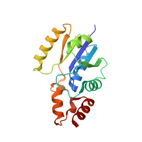
8I8I - PubMed Abstract:
Phosphopantetheine adenylyltransferase (EC. 2.7.7.3, PPAT) catalyzes the penultimate step of the multistep reaction in the coenzyme A (CoA) biosynthesis pathway. In this step, an adenylyl group from adenosine triphosphate (ATP) is transferred to 4'-phosphopantetheine (PNS) yielding 3'-dephospho-coenzyme A (dpCoA) and pyrophosphate (PP i ). PPAT from strain C3 of Klebsiella pneumoniae (KpPPAT) was cloned, expressed and purified. It was crystallized using 0.1 M HEPES buffer and PEG10000 at pH 7.5. The crystals belonged to tetragonal space group P4 1 2 1 2 with cell dimensions of a = b = 72.82 Å and c = 200.37 Å. The structure was determined using the molecular replacement method and refined to values of 0.208 and 0.255 for R cryst and R free factors, respectively. The structure determination showed the presence of three crystallographically independent molecules A, B and C in the asymmetric unit. The molecules A and B are observed in the form of a dimer in the asymmetric unit while molecule C belongs to the second dimer whose partner is related by crystallographic twofold symmetry. The polypeptide chain of KpPPAT folds into a β/α structure. The conformations of the side chains of several residues in the substrate binding site in KpPPAT are significantly different from those reported in other PPATs. As a result, the modes of binding of substrates, phosphopantetheine (PNS) and adenosine triphosphate (ATP) differ considerably. The binding studies using fluorescence spectroscopy indicated a K D value of 3.45 × 10 -4 M for ATP which is significantly lower than the corresponding values reported for PPAT from other species.
- Department of Biophysics, All India Institute of Medical Sciences, Ansari Nagar, New Delhi, 110029, India.
Organizational Affiliation: