8I4O

Design of a split green fluorescent protein for sensing and tracking an beta-amyloid


Experimental Data Snapshot

  • Method: X-RAY DIFFRACTION
  • Resolution: 3.10 Å
  • R-Value Free: 0.323 
  • R-Value Work: 0.252 
  • R-Value Observed: 0.256 

wwPDB Validation   3D Report Full Report


This is version 1.1 of the entry. See complete history


Literature

Engineering of a Fluorescent Protein for a Sensing of an Intrinsically Disordered Protein through Transition in the Chromophore State.

Yu, T.G.Lee, J.Yoon, J.Choi, J.M.Kim, D.G.Heo, W.D.Song, J.J.Kim, H.S.

(2023) JACS Au 3: 3055-3065

  • DOI: https://doi.org/10.1021/jacsau.3c00445
  • Primary Citation of Related Structures:  
    8I4O

  • PubMed Abstract: 

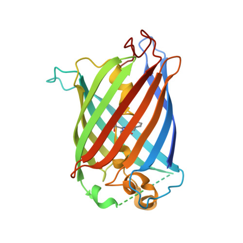
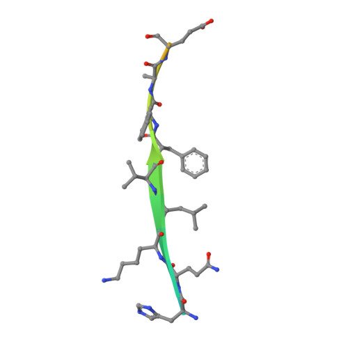
    Intrinsically disordered proteins (IDPs) not only play important roles in biological processes but are also linked with the pathogenesis of various human diseases. Specific and reliable sensing of IDPs is crucial for exploring their roles but remains elusive due to structural plasticity. Here, we present the development of a new type of fluorescent protein for the ratiometric sensing and tracking of an IDP. A β-strand of green fluorescent protein (GFP) was truncated, and the resulting GFP was further engineered to undergo the transition in the absorption maximum upon binding of a target motif within amyloid-β (Aβ) as a model IDP through rational design and directed evolution. Spectroscopic and structural analyses of the engineered truncated GFP demonstrated that a shift in the absorption maximum is driven by the change in the chromophore state from an anionic (460 nm) state into a neutral (390 nm) state as the Aβ binds, allowing a ratiometric detection of Aβ. The utility of the developed GFP was shown by the efficient and specific detection of an Aβ and the tracking of its conformational change and localization in astrocytes.


  • Organizational Affiliation

    Departement of Biological Sciences, Korea Advanced Institute of Science and Technology (KAIST), Daejeon 34141, Korea.


Macromolecules
Find similar proteins by:  (by identity cutoff)  |  3D Structure
Entity ID: 1
MoleculeChains Sequence LengthOrganismDetailsImage
Split Green flourescent protein
A, C, E, G, I
A, C, E, G, I, K
239Bacteria Latreille et al. 1825Mutation(s): 0 
Entity Groups  
Sequence Clusters30% Identity50% Identity70% Identity90% Identity95% Identity100% Identity
Sequence Annotations
Expand
  • Reference Sequence

Find similar proteins by:  Sequence   |   3D Structure  

Entity ID: 2
MoleculeChains Sequence LengthOrganismDetailsImage
Beta-amyloid
B, D, F, H, J
B, D, F, H, J, L
21Bacteria Latreille et al. 1825Mutation(s): 0 
UniProt & NIH Common Fund Data Resources
Find proteins for P05067 (Homo sapiens)
Explore P05067 
Go to UniProtKB:  P05067
PHAROS:  P05067
GTEx:  ENSG00000142192 
Entity Groups  
Sequence Clusters30% Identity50% Identity70% Identity90% Identity95% Identity100% Identity
UniProt GroupP05067
Sequence Annotations
Expand
  • Reference Sequence
Small Molecules
Modified Residues  1 Unique
IDChains TypeFormula2D DiagramParent
GYS
Query on GYS
A, C, E, G, I
A, C, E, G, I, K
L-PEPTIDE LINKINGC14 H15 N3 O5SER, TYR, GLY
Experimental Data & Validation

Experimental Data

  • Method: X-RAY DIFFRACTION
  • Resolution: 3.10 Å
  • R-Value Free: 0.323 
  • R-Value Work: 0.252 
  • R-Value Observed: 0.256 
  • Space Group: P 1
Unit Cell:
Length ( Å )Angle ( ˚ )
a = 64.804α = 98.38
b = 68.819β = 97.97
c = 101.239γ = 113.44
Software Package:
Software NamePurpose
PHENIXrefinement
HKL-2000data reduction
HKL-2000data scaling

Structure Validation

View Full Validation Report


Entry History & Funding Information

Deposition Data


Funding OrganizationLocationGrant Number
National Research Foundation (NRF, Korea)Korea, Republic Of2019R1I1A1A01040965
National Research Foundation (NRF, Korea)Korea, Republic Of2021R1A2C201421811

Revision History  (Full details and data files)

  • Version 1.0: 2023-11-29
    Type: Initial release
  • Version 1.1: 2023-12-13
    Changes: Database references