8HTE

Crystal structure of an effector mutant in complex with ubiquitin


Experimental Data Snapshot

  • Method: X-RAY DIFFRACTION
  • Resolution: 2.31 Å
  • R-Value Free: 0.228 
  • R-Value Work: 0.171 
  • R-Value Observed: 0.174 

wwPDB Validation   3D Report Full Report


Ligand Structure Quality Assessment 


This is version 1.1 of the entry. See complete history


Literature

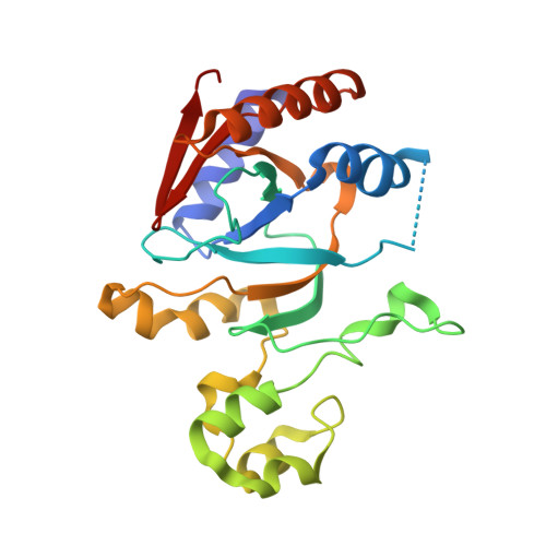
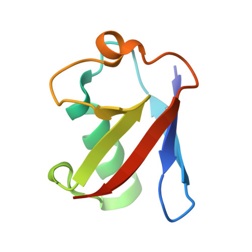
Molecular basis of threonine ADP-ribosylation of ubiquitin by bacterial ARTs.

Tan, J.Xu, Y.Wang, X.Yan, F.Xian, W.Liu, X.Chen, Y.Zhu, Y.Zhou, Y.

(2024) Nat Chem Biol 20: 463-472

  • DOI: https://doi.org/10.1038/s41589-023-01475-3
  • Primary Citation of Related Structures:  
    8HTC, 8HTD, 8HTE, 8HTF

  • PubMed Abstract: 

    Ubiquitination plays essential roles in eukaryotic cellular processes. The effector protein CteC from Chromobacterium violaceum blocks host ubiquitination by mono-ADP-ribosylation of ubiquitin (Ub) at residue T66. However, the structural basis for this modification is unknown. Here we report three crystal structures of CteC in complexes with Ub, NAD + or ADP-ribosylated Ub, which represent different catalytic states of CteC in the modification. CteC adopts a special 'D-E' catalytic motif for catalysis and binds NAD + in a half-ligand binding mode. The specific recognition of Ub by CteC is determined by a relatively separate Ub-targeting domain and a long loop L6, not the classic ADP-ribosylating turn-turn loop. Structural analyses with biochemical results reveal that CteC represents a large family of poly (ADP-ribose) polymerase (PARP)-like ADP-ribosyltransferases, which harbors chimeric features from the R-S-E and H-Y-E classes of ADP-ribosyltransferases. The family of CteC-like ADP-ribosyltransferases has a common 'D-E' catalytic consensus and exists extensively in bacteria and eukaryotic microorganisms.


  • Organizational Affiliation

    The MOE Key Laboratory for Biosystems Homeostasis & Protection and Innovation Center for Cell Signaling Network, Life Sciences Institute, Zhejiang University, Hangzhou, China.


Macromolecules
Find similar proteins by:  (by identity cutoff)  |  3D Structure
Entity ID: 1
MoleculeChains Sequence LengthOrganismDetailsImage
NAD(+)--protein-threonine ADP-ribosyltransferase233Chromobacterium violaceum ATCC 12472Mutation(s): 1 
Gene Names: cteCCV_1467
EC: 2.4.2
UniProt
Find proteins for Q7NY09 (Chromobacterium violaceum (strain ATCC 12472 / DSM 30191 / JCM 1249 / NBRC 12614 / NCIMB 9131 / NCTC 9757))
Explore Q7NY09 
Go to UniProtKB:  Q7NY09
Entity Groups  
Sequence Clusters30% Identity50% Identity70% Identity90% Identity95% Identity100% Identity
UniProt GroupQ7NY09
Sequence Annotations
Expand
  • Reference Sequence
Find similar proteins by:  (by identity cutoff)  |  3D Structure
Entity ID: 2
MoleculeChains Sequence LengthOrganismDetailsImage
Ubiquitin76Saccharomyces cerevisiae S288CMutation(s): 0 
Gene Names: RPL40AUBI1YIL148W
UniProt
Find proteins for P0CH08 (Saccharomyces cerevisiae (strain ATCC 204508 / S288c))
Explore P0CH08 
Go to UniProtKB:  P0CH08
Entity Groups  
Sequence Clusters30% Identity50% Identity70% Identity90% Identity95% Identity100% Identity
UniProt GroupP0CH08
Sequence Annotations
Expand
  • Reference Sequence
Small Molecules
Ligands 4 Unique
IDChains Name / Formula / InChI Key2D Diagram3D Interactions
AR6 (Subject of Investigation/LOI)
Query on AR6

Download Ideal Coordinates CCD File 
G [auth B][(2R,3S,4R,5R)-5-(6-AMINOPURIN-9-YL)-3,4-DIHYDROXY-OXOLAN-2-YL]METHYL [HYDROXY-[[(2R,3S,4R,5S)-3,4,5-TRIHYDROXYOXOLAN-2-YL]METHOXY]PHOSPHORYL] HYDROGEN PHOSPHATE
C15 H23 N5 O14 P2
SRNWOUGRCWSEMX-ZQSHOCFMSA-N
NCA (Subject of Investigation/LOI)
Query on NCA

Download Ideal Coordinates CCD File 
F [auth A]NICOTINAMIDE
C6 H6 N2 O
DFPAKSUCGFBDDF-UHFFFAOYSA-N
SO4
Query on SO4

Download Ideal Coordinates CCD File 
C [auth A],
D [auth A]
SULFATE ION
O4 S
QAOWNCQODCNURD-UHFFFAOYSA-L
GOL
Query on GOL

Download Ideal Coordinates CCD File 
E [auth A],
H [auth B]
GLYCEROL
C3 H8 O3
PEDCQBHIVMGVHV-UHFFFAOYSA-N
Experimental Data & Validation

Experimental Data

  • Method: X-RAY DIFFRACTION
  • Resolution: 2.31 Å
  • R-Value Free: 0.228 
  • R-Value Work: 0.171 
  • R-Value Observed: 0.174 
  • Space Group: P 21 21 21
Unit Cell:
Length ( Å )Angle ( ˚ )
a = 59.074α = 90
b = 70.478β = 90
c = 90.562γ = 90
Software Package:
Software NamePurpose
SCALEPACKdata scaling
PHENIXrefinement
PDB_EXTRACTdata extraction
HKL-2000data reduction
PHENIXphasing

Structure Validation

View Full Validation Report


Ligand Structure Quality Assessment 


Entry History & Funding Information

Deposition Data


Funding OrganizationLocationGrant Number
National Natural Science Foundation of China (NSFC)China81530068
National Natural Science Foundation of China (NSFC)China81322024
National Natural Science Foundation of China (NSFC)China31370722
National Natural Science Foundation of China (NSFC)China81561130162
National Natural Science Foundation of China (NSFC)China81501717
Ministry of Science and Technology (MoST, China)China2017YFA0503900

Revision History  (Full details and data files)

  • Version 1.0: 2023-11-22
    Type: Initial release
  • Version 1.1: 2024-04-10
    Changes: Database references