8HEB | pdb_00008heb

SARS-CoV-2 Spike trimer in complex with RmAb 9H1 Fab in the class 1 conformation


Experimental Data Snapshot

  • Method: ELECTRON MICROSCOPY
  • Resolution: 3.53 Å
  • Aggregation State: PARTICLE 
  • Reconstruction Method: SINGLE PARTICLE 

wwPDB Validation 3D Report Full Report

Validation slider image for 8HEB

This is version 1.2 of the entry. See complete history

Literature

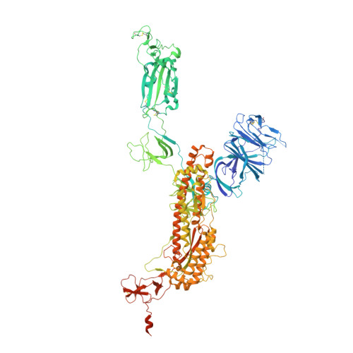
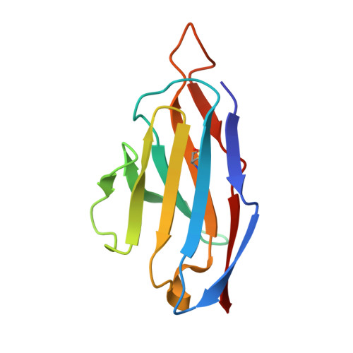
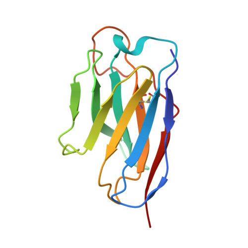
Mechanism of an RBM-targeted rabbit monoclonal antibody 9H1 neutralizing SARS-CoV-2.

Chu, X.Ding, X.Yang, Y.Lu, Y.Li, T.Gao, Y.Zheng, L.Xiao, H.Yang, T.Cheng, H.Huang, H.Liu, Y.Lou, Y.Wu, C.Chen, Y.Yang, H.Ji, X.Guo, H.

(2023) Biochem Biophys Res Commun 660: 43-49

  • DOI: https://doi.org/10.1016/j.bbrc.2023.04.002
  • Primary Citation Related Structures: 
    8HEB, 8HEC, 8HED

  • PubMed Abstract: 

    The COVID-19 pandemic, caused by SARS-CoV-2, has led to over 750 million infections and 6.8 million deaths worldwide since late 2019. Due to the continuous evolution of SARS-CoV-2, many significant variants have emerged, creating ongoing challenges to the prevention and treatment of the pandemic. Therefore, the study of antibody responses against SARS-CoV-2 is essential for the development of vaccines and therapeutics. Here we perform single particle cryo-electron microscopy (cryo-EM) structure determination of a rabbit monoclonal antibody (RmAb) 9H1 in complex with the SARS-CoV-2 wild-type (WT) spike trimer. Our structural analysis shows that 9H1 interacts with the receptor-binding motif (RBM) region of the receptor-binding domain (RBD) on the spike protein and by directly competing with angiotensin-converting enzyme 2 (ACE2), it blocks the binding of the virus to the receptor and achieves neutralization. Our findings suggest that utilizing rabbit-derived mAbs provides valuable insights into the molecular interactions between neutralizing antibodies and spike proteins and may also facilitate the development of therapeutic antibodies and expand the antibody library.


  • Organizational Affiliation
    • The State Key Laboratory of Pharmaceutical Biotechnology, School of Life Sciences, Nanjing University, Nanjing, Jiangsu, 210023, China.

Macromolecule Content 

  • Total Structure Weight: 487.5 kDa 
  • Atom Count: 29,912 
  • Modeled Residue Count: 3,769 
  • Deposited Residue Count: 4,305 
  • Unique protein chains: 3

Macromolecules

Find similar proteins by:|  3D Structure
Entity ID: 1
MoleculeChains  Sequence LengthOrganismDetailsImage
Spike glycoprotein
A, B, C
1,208Severe acute respiratory syndrome coronavirus 2Mutation(s): 5 
Gene Names: S2
UniProt
Find proteins for P0DTC2 (Severe acute respiratory syndrome coronavirus 2)
Explore P0DTC2 
Go to UniProtKB:  P0DTC2
Entity Groups
Sequence Clusters30% Identity50% Identity70% Identity90% Identity95% Identity100% Identity
UniProt GroupP0DTC2
Glycosylation
Glycosylation Sites: 15Go to GlyGen: P0DTC2-1
Sequence Annotations
Expand
Reference Sequence
Find similar proteins by:|  3D Structure
Entity ID: 2
MoleculeChains  Sequence LengthOrganismDetailsImage
rabbit antibody 9H1 light chain
D, F, H
110Oryctolagus cuniculusMutation(s): 0 
Find similar proteins by:|  3D Structure
Entity ID: 3
MoleculeChains  Sequence LengthOrganismDetailsImage
rabbit antibody 9H1 heavy chain
E, G, I
117Oryctolagus cuniculusMutation(s): 0 

Small Molecules

Ligands 1 Unique
IDChains Name / Formula / InChI Key2D Diagram3D Interactions
NAG
(Subject of Investigation/LOI)

Query on NAG


Download:Ideal Coordinates CCD File
AB [auth C]
BA [auth A]
BB [auth C]
CA [auth A]
DA [auth A]
AB [auth C],
BA [auth A],
BB [auth C],
CA [auth A],
DA [auth A],
EA [auth A],
FA [auth A],
GA [auth A],
HA [auth A],
IA [auth A],
JA [auth A],
KA [auth B],
LA [auth B],
MA [auth B],
NA [auth B],
OA [auth B],
PA [auth B],
QA [auth B],
RA [auth B],
SA [auth B],
TA [auth C],
UA [auth C],
VA [auth C],
WA [auth C],
XA [auth C],
YA [auth C],
ZA [auth C]
2-acetamido-2-deoxy-beta-D-glucopyranose
C8 H15 N O6
OVRNDRQMDRJTHS-FMDGEEDCSA-N

Experimental Data & Validation

Experimental Data

  • Method: ELECTRON MICROSCOPY
  • Resolution: 3.53 Å
  • Aggregation State: PARTICLE 
  • Reconstruction Method: SINGLE PARTICLE 
EM Software:
TaskSoftware PackageVersion
MODEL REFINEMENTPHENIX

Structure Validation

View Full Validation Report


Entry History 

& Funding Information

Deposition Data


Funding OrganizationLocationGrant Number
Other governmentChina--

Revision History  (Full details and data files)

  • Version 1.0: 2023-04-26
    Type: Initial release
  • Version 1.1: 2024-10-30
    Changes: Data collection, Structure summary
  • Version 1.2: 2025-06-25
    Changes: Data collection