8H9Y

Crystal structure of voltage-gated sodium channel NavAb N49K/L176Q mutant in calcium ion condition


Experimental Data Snapshot

  • Method: X-RAY DIFFRACTION
  • Resolution: 3.40 Å
  • R-Value Free: 0.264 
  • R-Value Work: 0.254 
  • R-Value Observed: 0.255 

wwPDB Validation   3D Report Full Report


This is version 1.0 of the entry. See complete history


Literature

The structural basis of divalent cation block in a tetrameric prokaryotic sodium channel.

Irie, K.Oda, Y.Sumikama, T.Oshima, A.Fujiyoshi, Y.

(2023) Nat Commun 14: 4236-4236

  • DOI: https://doi.org/10.1038/s41467-023-39987-0
  • Primary Citation of Related Structures:  
    8H9O, 8H9W, 8H9X, 8H9Y, 8HA1, 8HA2

  • PubMed Abstract: 

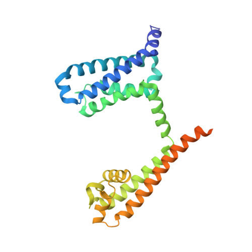
    Divalent cation block is observed in various tetrameric ion channels. For blocking, a divalent cation is thought to bind in the ion pathway of the channel, but such block has not yet been directly observed. So, the behaviour of these blocking divalent cations remains still uncertain. Here, we elucidated the mechanism of the divalent cation block by reproducing the blocking effect into NavAb, a well-studied tetrameric sodium channel. Our crystal structures of NavAb mutants show that the mutations increasing the hydrophilicity of the inner vestibule of the pore domain enable a divalent cation to stack on the ion pathway. Furthermore, non-equilibrium molecular dynamics simulation showed that the stacking calcium ion repel sodium ion at the bottom of the selectivity filter. These results suggest the primary process of the divalent cation block mechanism in tetrameric cation channels.


  • Organizational Affiliation

    Department of Biophysical Chemistry, School of Pharmaceutical Sciences, Wakayama Medical University, 25-1, Shichibancho, Wakayama, 640-8156, Japan. kirie@wakayama-med.ac.jp.


Macromolecules
Find similar proteins by:  (by identity cutoff)  |  3D Structure
Entity ID: 1
MoleculeChains Sequence LengthOrganismDetailsImage
Ion transport protein271Aliarcobacter butzleriMutation(s): 2 
Gene Names: Abu_1752
Membrane Entity: Yes 
UniProt
Find proteins for A8EVM5 (Aliarcobacter butzleri (strain RM4018))
Explore A8EVM5 
Go to UniProtKB:  A8EVM5
Entity Groups  
Sequence Clusters30% Identity50% Identity70% Identity90% Identity95% Identity100% Identity
UniProt GroupA8EVM5
Sequence Annotations
Expand
  • Reference Sequence
Small Molecules
Ligands 4 Unique
IDChains Name / Formula / InChI Key2D Diagram3D Interactions
PX4
Query on PX4

Download Ideal Coordinates CCD File 
E [auth A]
F [auth A]
G [auth A]
H [auth A]
I [auth A]
E [auth A],
F [auth A],
G [auth A],
H [auth A],
I [auth A],
J [auth A],
K [auth A],
L [auth A]
1,2-DIMYRISTOYL-SN-GLYCERO-3-PHOSPHOCHOLINE
C36 H73 N O8 P
CITHEXJVPOWHKC-UUWRZZSWSA-O
1N7
Query on 1N7

Download Ideal Coordinates CCD File 
D [auth A]CHAPSO
C32 H59 N2 O8 S
GUQQBLRVXOUDTN-XOHPMCGNSA-O
LMT
Query on LMT

Download Ideal Coordinates CCD File 
B [auth A],
C [auth A]
DODECYL-BETA-D-MALTOSIDE
C24 H46 O11
NLEBIOOXCVAHBD-QKMCSOCLSA-N
CA (Subject of Investigation/LOI)
Query on CA

Download Ideal Coordinates CCD File 
M [auth A],
N [auth A]
CALCIUM ION
Ca
BHPQYMZQTOCNFJ-UHFFFAOYSA-N
Experimental Data & Validation

Experimental Data

  • Method: X-RAY DIFFRACTION
  • Resolution: 3.40 Å
  • R-Value Free: 0.264 
  • R-Value Work: 0.254 
  • R-Value Observed: 0.255 
  • Space Group: I 4 2 2
Unit Cell:
Length ( Å )Angle ( ˚ )
a = 127.94α = 90
b = 127.94β = 90
c = 201.92γ = 90
Software Package:
Software NamePurpose
PHENIXrefinement
XDSdata reduction
XSCALEdata scaling
PHASERphasing

Structure Validation

View Full Validation Report


Entry History & Funding Information

Deposition Data


Funding OrganizationLocationGrant Number
Japan Society for the Promotion of Science (JSPS)Japan17K17795
Japan Society for the Promotion of Science (JSPS)Japan20K09193

Revision History  (Full details and data files)

  • Version 1.0: 2023-07-26
    Type: Initial release