8H1P

Cryo-EM structure of the human RAD52 protein


Experimental Data Snapshot

  • Method: ELECTRON MICROSCOPY
  • Resolution: 3.48 Å
  • Aggregation State: PARTICLE 
  • Reconstruction Method: SINGLE PARTICLE 

wwPDB Validation   3D Report Full Report


This is version 1.1 of the entry. See complete history


Literature

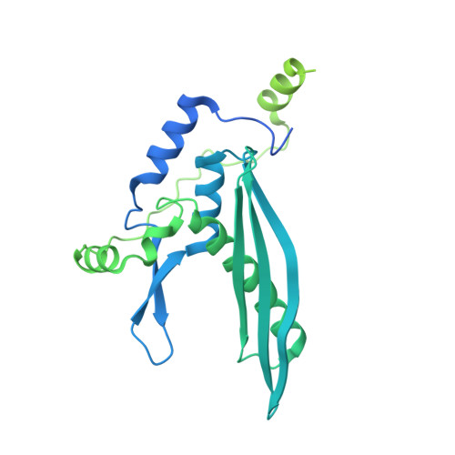
The cryo-EM structure of full-length RAD52 protein contains an undecameric ring.

Kinoshita, C.Takizawa, Y.Saotome, M.Ogino, S.Kurumizaka, H.Kagawa, W.

(2023) FEBS Open Bio 13: 408-418

  • DOI: https://doi.org/10.1002/2211-5463.13565
  • Primary Citation of Related Structures:  
    8H1P

  • PubMed Abstract: 

    The human RAD52 protein, which forms an oligomeric ring structure, is involved in DNA double-strand break repair. The N-terminal half of RAD52 is primarily responsible for self-oligomerisation and DNA binding. Crystallographic studies have revealed the detailed structure of the N-terminal half. However, only low-resolution structures have been reported for the full-length protein, and thus the structural role of the C-terminal half in self-oligomerisation has remained elusive. In this study, we determined the solution structure of the human RAD52 protein by cryo-electron microscopy (cryo-EM), at an average resolution of 3.5 Å. The structure revealed an undecameric ring that is nearly identical to the crystal structures of the N-terminal half. The cryo-EM map for the C-terminal half was poorly defined, indicating that the region is intrinsically disordered. The present cryo-EM structure provides important insights into the mechanistic roles played by the N-terminal and C-terminal halves of RAD52 during DNA double-strand break repair.


  • Organizational Affiliation

    Department of Chemistry, Graduate School of Science and Engineering, Meisei University, Tokyo, Japan.


Macromolecules
Find similar proteins by:  (by identity cutoff)  |  3D Structure
Entity ID: 1
MoleculeChains Sequence LengthOrganismDetailsImage
DNA repair protein RAD52 homolog
A, B, C, D, E
A, B, C, D, E, F, G, H, I, J, K
421Homo sapiensMutation(s): 0 
Gene Names: RAD52
UniProt & NIH Common Fund Data Resources
Find proteins for P43351 (Homo sapiens)
Explore P43351 
Go to UniProtKB:  P43351
PHAROS:  P43351
GTEx:  ENSG00000002016 
Entity Groups  
Sequence Clusters30% Identity50% Identity70% Identity90% Identity95% Identity100% Identity
UniProt GroupP43351
Sequence Annotations
Expand
  • Reference Sequence
Experimental Data & Validation

Experimental Data

  • Method: ELECTRON MICROSCOPY
  • Resolution: 3.48 Å
  • Aggregation State: PARTICLE 
  • Reconstruction Method: SINGLE PARTICLE 
EM Software:
TaskSoftware PackageVersion
RECONSTRUCTIONRELION3.1

Structure Validation

View Full Validation Report


Entry History & Funding Information

Deposition Data


Funding OrganizationLocationGrant Number
Japan Society for the Promotion of Science (JSPS)JapanJP18H05534
Japan Society for the Promotion of Science (JSPS)Japan19K12328
Japan Society for the Promotion of Science (JSPS)Japan20H00449
Japan Society for the Promotion of Science (JSPS)Japan22H03743
Japan Society for the Promotion of Science (JSPS)Japan22K06098
Japan Science and TechnologyJapanJPMJER1901
Japan Agency for Medical Research and Development (AMED)JapanJP22am121009

Revision History  (Full details and data files)

  • Version 1.0: 2023-02-08
    Type: Initial release
  • Version 1.1: 2023-03-22
    Changes: Database references