Biodegradation of highly crystallized poly(ethylene terephthalate) through cell surface codisplay of bacterial PETase and hydrophobin.
Chen, Z., Duan, R., Xiao, Y., Wei, Y., Zhang, H., Sun, X., Wang, S., Cheng, Y., Wang, X., Tong, S., Yao, Y., Zhu, C., Yang, H., Wang, Y., Wang, Z.(2022) Nat Commun 13: 7138-7138
- PubMed: 36414665 Search on PubMedSearch on PubMed Central
- DOI: https://doi.org/10.1038/s41467-022-34908-z
- Primary Citation Related Structures:
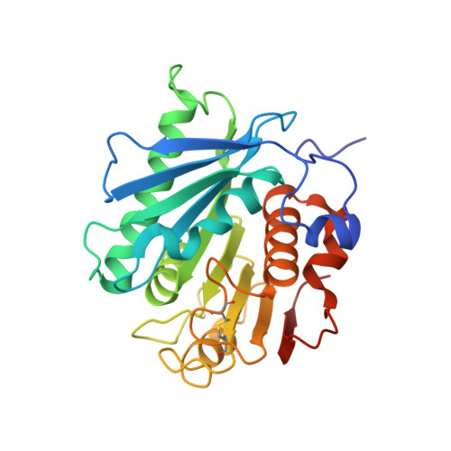
8GU4, 8GU5 - PubMed Abstract:
The process of recycling poly(ethylene terephthalate) (PET) remains a major challenge due to the enzymatic degradation of high-crystallinity PET (hcPET). Recently, a bacterial PET-degrading enzyme, PETase, was found to have the ability to degrade the hcPET, but with low enzymatic activity. Here we present an engineered whole-cell biocatalyst to simulate both the adsorption and degradation steps in the enzymatic degradation process of PETase to achieve the efficient degradation of hcPET. Our data shows that the adhesive unit hydrophobin and degradation unit PETase are functionally displayed on the surface of yeast cells. The turnover rate of the whole-cell biocatalyst toward hcPET (crystallinity of 45%) dramatically increases approximately 328.8-fold compared with that of purified PETase at 30 °C. In addition, molecular dynamics simulations explain how the enhanced adhesion can promote the enzymatic degradation of PET. This study demonstrates engineering the whole-cell catalyst is an efficient strategy for biodegradation of PET.
- School of Life Sciences, Tianjin Key Laboratory of Function and Application of Biological Macromolecular Structures, College of Precision Instrument and Opto-electronics Engineering, Key Laboratory of Systems Bioengineering (Ministry of Education), Tianjin University, Tianjin, 300072, China.
Organizational Affiliation: