8GQR

Crystal structure of VioD with FAD


Experimental Data Snapshot

  • Method: X-RAY DIFFRACTION
  • Resolution: 2.10 Å
  • R-Value Free: 0.226 
  • R-Value Work: 0.190 

Starting Model: experimental
View more details

wwPDB Validation   3D Report Full Report


Ligand Structure Quality Assessment 


This is version 1.2 of the entry. See complete history


Literature

Structural basis for substrate binding and catalytic mechanism of the key enzyme VioD in the violacein synthesis pathway.

Xu, M.Xu, D.Gao, M.Zhuang, X.Wang, W.Sun, B.Ran, T.

(2023) Proteins 91: 956-966

  • DOI: https://doi.org/10.1002/prot.26484
  • Primary Citation of Related Structures:  
    8GQR, 8H0M

  • PubMed Abstract: 

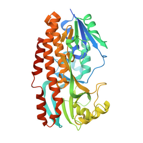
    Violacein is a pigment synthesized by gram-negative bacteria with various biological activities such as antimicrobial, antiviral, and anticancer activities. VioD is a key oxygenase converting protodeoxyviolaceinic acid to protoviolaceinic acid in violacein biosynthesis. To elucidate the catalytic mechanism of VioD, here, we resolved two crystal structures of VioD, a binary complex structure containing VioD and a FAD and a ternary complex structure composed of VioD, a FAD and a 2-ethyl-1-hexanol (EHN). Structural analysis revealed a deep funnel like binding pocket with wide entrance, this pocket is positively charged. The EHN is located at the deep bottom of the binding pocket near isoalloxazine ring. Further docking simulation help us to propose the mechanism of the hydroxylation of the substrate catalyzed by VioD. Bioinformatic analysis suggested and emphasized the importance of the conserved residues involved in substrate binding. Our results provide a structural basis for the catalytic mechanism of VioD.


  • Organizational Affiliation

    Department of Microbiology, College of Life Sciences, Nanjing Agricultural University, Nanjing, China.


Macromolecules
Find similar proteins by:  (by identity cutoff)  |  3D Structure
Entity ID: 1
MoleculeChains Sequence LengthOrganismDetailsImage
VioD
A, B
403Duganella sp. ZLP-XIMutation(s): 0 
UniProt
Find proteins for A0A024AX32 (Duganella sp. ZLP-XI)
Explore A0A024AX32 
Go to UniProtKB:  A0A024AX32
Entity Groups  
Sequence Clusters30% Identity50% Identity70% Identity90% Identity95% Identity100% Identity
UniProt GroupA0A024AX32
Sequence Annotations
Expand
  • Reference Sequence
Small Molecules
Ligands 3 Unique
IDChains Name / Formula / InChI Key2D Diagram3D Interactions
FAD (Subject of Investigation/LOI)
Query on FAD

Download Ideal Coordinates CCD File 
G [auth A],
K [auth B]
FLAVIN-ADENINE DINUCLEOTIDE
C27 H33 N9 O15 P2
VWWQXMAJTJZDQX-UYBVJOGSSA-N
SO4
Query on SO4

Download Ideal Coordinates CCD File 
C [auth A]
D [auth A]
E [auth A]
H [auth B]
I [auth B]
C [auth A],
D [auth A],
E [auth A],
H [auth B],
I [auth B],
J [auth B]
SULFATE ION
O4 S
QAOWNCQODCNURD-UHFFFAOYSA-L
GOL
Query on GOL

Download Ideal Coordinates CCD File 
F [auth A]GLYCEROL
C3 H8 O3
PEDCQBHIVMGVHV-UHFFFAOYSA-N
Experimental Data & Validation

Experimental Data

  • Method: X-RAY DIFFRACTION
  • Resolution: 2.10 Å
  • R-Value Free: 0.226 
  • R-Value Work: 0.190 
  • Space Group: P 21 21 21
Unit Cell:
Length ( Å )Angle ( ˚ )
a = 50.603α = 90
b = 100.869β = 90
c = 162.815γ = 90
Software Package:
Software NamePurpose
PHENIXrefinement
XDSdata reduction
SCALAdata scaling
PHASERphasing

Structure Validation

View Full Validation Report


Ligand Structure Quality Assessment 


Entry History & Funding Information

Deposition Data


Funding OrganizationLocationGrant Number
National Natural Science Foundation of China (NSFC)China31770074

Revision History  (Full details and data files)

  • Version 1.0: 2023-03-29
    Type: Initial release
  • Version 1.1: 2023-06-21
    Changes: Database references
  • Version 1.2: 2023-11-29
    Changes: Data collection, Refinement description