8GM2

Crystal structure of T252E-CYP199A4 in complex with 4-methoxybenzoic acid soaked with 2 mM hydrogen peroxide


Experimental Data Snapshot

  • Method: X-RAY DIFFRACTION
  • Resolution: 2.33 Å
  • R-Value Free: 0.269 
  • R-Value Work: 0.208 
  • R-Value Observed: 0.214 

wwPDB Validation   3D Report Full Report


Ligand Structure Quality Assessment 


This is version 1.1 of the entry. See complete history


Literature

An In Crystallo Reaction with an Engineered Cytochrome P450 Peroxygenase.

Lee, J.H.Z.Bruning, J.B.Bell, S.G.

(2024) Chemistry 30: e202303335-e202303335

  • DOI: https://doi.org/10.1002/chem.202303335
  • Primary Citation of Related Structures:  
    8GLY, 8GLZ, 8GM1, 8GM2

  • PubMed Abstract: 

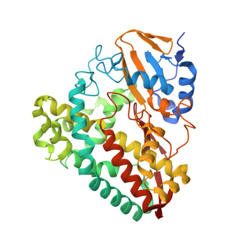
    The cytochrome P450 monooxygenases (CYPs) are a class of heme-thiolate enzymes that insert oxygen into unactivated C-H bonds. These enzymes can be converted into peroxygenases via protein engineering, which enables their activity to occur using hydrogen peroxide (H 2 O 2 ) without the requirement for additional nicotinamide co-factors or partner proteins. Here, we demonstrate that soaking crystals of an engineered P450 peroxygenase with H 2 O 2 enables the enzymatic reaction to occur within the crystal. Crystals of the designed P450 peroxygenase, the T252E mutant of CYP199A4, in complex with 4-methoxybenzoic acid were soaked with different concentrations of H 2 O 2 for varying times to initiate the in crystallo O-demethylation reaction. Crystal structures of T252E-CYP199A4 showed a distinct loss of electron density that was consistent with the O-demethylated metabolite, 4-hydroxybenzoic acid. A new X-ray crystal structure of this enzyme with the 4-hydroxybenzoic acid product was obtained to enable comparison alongside the existing substrate-bound structure. The visualisation of enzymatic catalysis in action is challenging in structural biology and the ability to initiate the reactions of P450 enzymes, in crystallo by simply soaking crystals with H 2 O 2 will enable new structural biology methods and techniques to be applied to study their mechanism of action.


  • Organizational Affiliation

    Department of Chemistry, University of Adelaide, Adelaide, SA 5005, Australia.


Macromolecules
Find similar proteins by:  (by identity cutoff)  |  3D Structure
Entity ID: 1
MoleculeChains Sequence LengthOrganismDetailsImage
Cytochrome P450410Rhodopseudomonas palustris HaA2Mutation(s): 1 
Gene Names: RPB_3613
UniProt
Find proteins for Q2IU02 (Rhodopseudomonas palustris (strain HaA2))
Explore Q2IU02 
Go to UniProtKB:  Q2IU02
Entity Groups  
Sequence Clusters30% Identity50% Identity70% Identity90% Identity95% Identity100% Identity
UniProt GroupQ2IU02
Sequence Annotations
Expand
  • Reference Sequence
Small Molecules
Ligands 2 Unique
IDChains Name / Formula / InChI Key2D Diagram3D Interactions
HEM
Query on HEM

Download Ideal Coordinates CCD File 
C [auth A]PROTOPORPHYRIN IX CONTAINING FE
C34 H32 Fe N4 O4
KABFMIBPWCXCRK-RGGAHWMASA-L
ANN (Subject of Investigation/LOI)
Query on ANN

Download Ideal Coordinates CCD File 
B [auth A]4-METHOXYBENZOIC ACID
C8 H8 O3
ZEYHEAKUIGZSGI-UHFFFAOYSA-N
Experimental Data & Validation

Experimental Data

  • Method: X-RAY DIFFRACTION
  • Resolution: 2.33 Å
  • R-Value Free: 0.269 
  • R-Value Work: 0.208 
  • R-Value Observed: 0.214 
  • Space Group: P 1 21 1
Unit Cell:
Length ( Å )Angle ( ˚ )
a = 44.372α = 90
b = 51.64β = 92.31
c = 79.055γ = 90
Software Package:
Software NamePurpose
PHENIXrefinement
Aimlessdata scaling
iMOSFLMdata reduction
PHASERphasing

Structure Validation

View Full Validation Report


Ligand Structure Quality Assessment 


Entry History & Funding Information

Deposition Data


Funding OrganizationLocationGrant Number
Australian Research Council (ARC)AustraliaDP140103229

Revision History  (Full details and data files)

  • Version 1.0: 2023-12-27
    Type: Initial release
  • Version 1.1: 2024-02-21
    Changes: Database references