8GLF

Crystal Structure of Human CD1b in Complex with Sphingomyelin C34:2


Experimental Data Snapshot

  • Method: X-RAY DIFFRACTION
  • Resolution: 2.00 Å
  • R-Value Free: 0.231 
  • R-Value Work: 0.176 
  • R-Value Observed: 0.178 

wwPDB Validation   3D Report Full Report


Ligand Structure Quality Assessment 


This is version 1.1 of the entry. See complete history


Literature

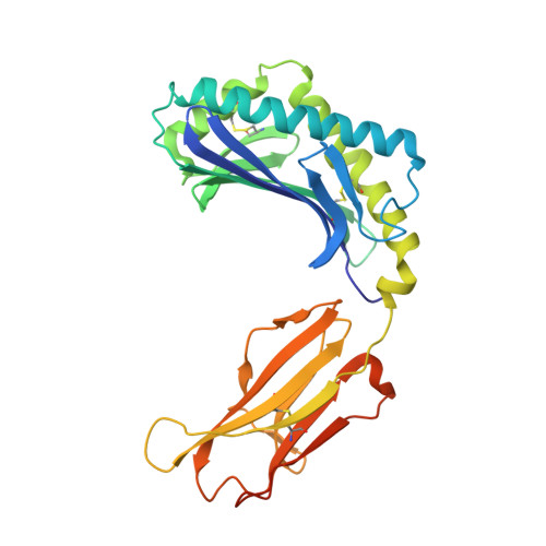
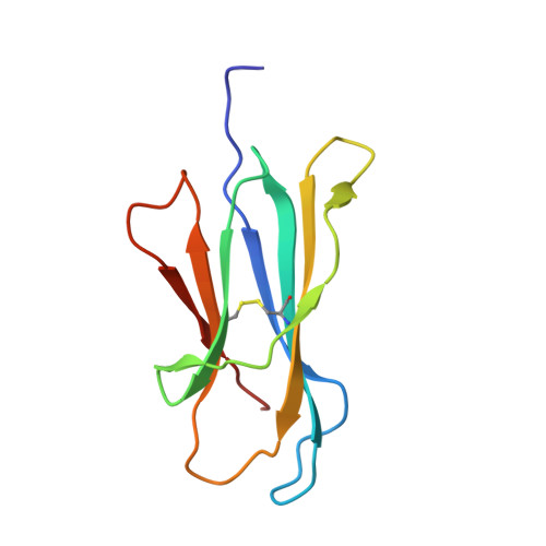
CD1 lipidomes reveal lipid-binding motifs and size-based antigen-display mechanisms.

Huang, S.Shahine, A.Cheng, T.Y.Chen, Y.L.Ng, S.W.Balaji, G.R.Farquhar, R.Gras, S.Hardman, C.S.Altman, J.D.Tahiri, N.Minnaard, A.J.Ogg, G.S.Mayfield, J.A.Rossjohn, J.Moody, D.B.

(2023) Cell 186: 4583-4596.e13

  • DOI: https://doi.org/10.1016/j.cell.2023.08.022
  • Primary Citation of Related Structures:  
    8GLE, 8GLF, 8GLG, 8GLH, 8GLI

  • PubMed Abstract: 

    The CD1 system binds lipid antigens for display to T cells. Here, we solved lipidomes for the four human CD1 antigen-presenting molecules, providing a map of self-lipid display. Answering a basic question, the detection of >2,000 CD1-lipid complexes demonstrates broad presentation of self-sphingolipids and phospholipids. Whereas peptide antigens are chemically processed, many lipids are presented in an unaltered form. However, each type of CD1 protein differentially edits the self-lipidome to show distinct capture motifs based on lipid length and chemical composition, suggesting general antigen display mechanisms. For CD1a and CD1d, lipid size matches the CD1 cleft volume. CD1c cleft size is more variable, and CD1b is the outlier, where ligands and clefts show an extreme size mismatch that is explained by uniformly seating two small lipids in one cleft. Furthermore, the list of compounds that comprise the integrated CD1 lipidome supports the ongoing discovery of lipid blockers and antigens for T cells.


  • Organizational Affiliation

    Division of Rheumatology, Immunity and Inflammation, Brigham and Women's Hospital, Harvard Medical School, Boston, MA 02115, USA. Electronic address: shouxiong.huang@uc.edu.


Macromolecules
Find similar proteins by:  (by identity cutoff)  |  3D Structure
Entity ID: 1
MoleculeChains Sequence LengthOrganismDetailsImage
T-cell surface glycoprotein CD1b300Homo sapiensMutation(s): 0 
Gene Names: CD1B
UniProt & NIH Common Fund Data Resources
Find proteins for P29016 (Homo sapiens)
Explore P29016 
Go to UniProtKB:  P29016
PHAROS:  P29016
GTEx:  ENSG00000158485 
Entity Groups  
Sequence Clusters30% Identity50% Identity70% Identity90% Identity95% Identity100% Identity
UniProt GroupP29016
Sequence Annotations
Expand
  • Reference Sequence
Find similar proteins by:  (by identity cutoff)  |  3D Structure
Entity ID: 2
MoleculeChains Sequence LengthOrganismDetailsImage
Beta-2-microglobulin101Homo sapiensMutation(s): 0 
Gene Names: B2MCDABP0092HDCMA22P
UniProt & NIH Common Fund Data Resources
Find proteins for P61769 (Homo sapiens)
Explore P61769 
Go to UniProtKB:  P61769
PHAROS:  P61769
GTEx:  ENSG00000166710 
Entity Groups  
Sequence Clusters30% Identity50% Identity70% Identity90% Identity95% Identity100% Identity
UniProt GroupP61769
Sequence Annotations
Expand
  • Reference Sequence
Oligosaccharides

Help

Entity ID: 3
MoleculeChains Length2D Diagram Glycosylation3D Interactions
2-acetamido-2-deoxy-beta-D-glucopyranose-(1-4)-2-acetamido-2-deoxy-beta-D-glucopyranoseC [auth D]2N-Glycosylation
Glycosylation Resources
GlyTouCan:  G42666HT
GlyCosmos:  G42666HT
GlyGen:  G42666HT
Entity ID: 4
MoleculeChains Length2D Diagram Glycosylation3D Interactions
alpha-D-mannopyranose-(1-3)-[alpha-D-mannopyranose-(1-6)]beta-D-mannopyranose-(1-4)-2-acetamido-2-deoxy-beta-D-glucopyranose-(1-4)-[alpha-L-fucopyranose-(1-3)][alpha-L-fucopyranose-(1-6)]2-acetamido-2-deoxy-beta-D-glucopyranoseD [auth C]7N-Glycosylation
Glycosylation Resources
GlyTouCan:  G11460AB
GlyCosmos:  G11460AB
GlyGen:  G11460AB
Small Molecules
Ligands 6 Unique
IDChains Name / Formula / InChI Key2D Diagram3D Interactions
3LY (Subject of Investigation/LOI)
Query on 3LY

Download Ideal Coordinates CCD File 
G [auth A][(~{Z},2~{R},3~{S})-2-(hexadecanoylamino)-3-oxidanyl-octadec-4-enyl] 2-(trimethyl-$l^{4}-azanyl)ethyl hydrogen phosphate
C39 H80 N2 O6 P
RWKUXQNLWDTSLO-OPXWDTKOSA-O
6UL (Subject of Investigation/LOI)
Query on 6UL

Download Ideal Coordinates CCD File 
H [auth A]TETRACOSYL PALMITATE
C40 H80 O2
KKTAIMZGSAQXRE-UHFFFAOYSA-N
IOD
Query on IOD

Download Ideal Coordinates CCD File 
M [auth A],
N [auth B]
IODIDE ION
I
XMBWDFGMSWQBCA-UHFFFAOYSA-M
EDO
Query on EDO

Download Ideal Coordinates CCD File 
E [auth A],
F [auth A]
1,2-ETHANEDIOL
C2 H6 O2
LYCAIKOWRPUZTN-UHFFFAOYSA-N
NI
Query on NI

Download Ideal Coordinates CCD File 
O [auth B]NICKEL (II) ION
Ni
VEQPNABPJHWNSG-UHFFFAOYSA-N
CL
Query on CL

Download Ideal Coordinates CCD File 
I [auth A],
J [auth A],
K [auth A],
L [auth A]
CHLORIDE ION
Cl
VEXZGXHMUGYJMC-UHFFFAOYSA-M
Experimental Data & Validation

Experimental Data

  • Method: X-RAY DIFFRACTION
  • Resolution: 2.00 Å
  • R-Value Free: 0.231 
  • R-Value Work: 0.176 
  • R-Value Observed: 0.178 
  • Space Group: P 21 21 21
Unit Cell:
Length ( Å )Angle ( ˚ )
a = 57.57α = 90
b = 77.98β = 90
c = 91.32γ = 90
Software Package:
Software NamePurpose
MOSFLMdata reduction
Aimlessdata scaling
PHENIXphasing
PHENIXrefinement

Structure Validation

View Full Validation Report


Ligand Structure Quality Assessment 


Entry History & Funding Information

Deposition Data

  • Released Date: 2023-09-20 
  • Deposition Author(s): Shahine, A.

Funding OrganizationLocationGrant Number
Australian Research Council (ARC)AustraliaDE210101031

Revision History  (Full details and data files)

  • Version 1.0: 2023-09-20
    Type: Initial release
  • Version 1.1: 2024-05-01
    Changes: Database references