Structure and function of a hexameric cyanophycin synthetase 2.
Markus, L.M.D., Sharon, I., Munro, K., Grogg, M., Hilvert, D., Strauss, M., Schmeing, T.M.(2023) Protein Sci 32: e4685-e4685
- PubMed: 37222490 Search on PubMedSearch on PubMed Central
- DOI: https://doi.org/10.1002/pro.4685
- Primary Citation Related Structures:
8FXH, 8FXI - PubMed Abstract:
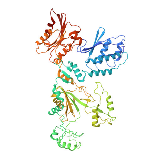
Cyanophycin is a natural polymer composed of a poly-aspartate backbone with arginine attached to each of the aspartate sidechains. Produced by a wide range of bacteria, which mainly use it as a store of fixed nitrogen, it has many promising industrial applications. Cyanophycin can be synthesized from the amino acids Asp and Arg by the widespread cyanophycin synthetase 1 (CphA1), or from the dipeptide β-Asp-Arg by the cyanobacterial enzyme cyanophycin synthetase 2 (CphA2). CphA2 enzymes display a range of oligomeric states, from dimers to dodecamers. Recently, the crystal structure of a CphA2 dimer was solved but could not be obtained in complex with substrate. Here, we report cryo-EM structures of the hexameric CphA2 from Stanieria sp. at ~2.8 Å resolution, both with and without ATP analog and cyanophycin. The structures show a two-fold symmetrical, trimer-of-dimers hexameric architecture, and substrate-binding interactions that are similar to those of CphA1. Mutagenesis experiments demonstrate the importance of several conserved substrate-binding residues. We also find that a Q416A/R528G double mutation prevents hexamer formation and use this double mutant to show that hexamerization augments the rate of cyanophycin synthesis. Together, these results increase our mechanistic understanding of how an interesting green polymer is biosynthesized.
- Department of Biochemistry, McGill University, Montréal, Quebec, Canada.
Organizational Affiliation: