8FWH | pdb_00008fwh

Crystal structure of bivalent antibody Fab fragment of Anti-human LAG3 (22D2)


Experimental Data Snapshot

  • Method: X-RAY DIFFRACTION
  • Resolution: 2.83 Å
  • R-Value Free: 
    0.277 (Depositor), 0.273 (DCC) 
  • R-Value Work: 
    0.216 (Depositor), 0.217 (DCC) 

Starting Model: experimental
View more details

wwPDB Validation 3D Report Full Report

Validation slider image for 8FWH

Ligand Structure Quality Assessment 


This is version 1.2 of the entry. See complete history

Literature

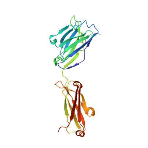
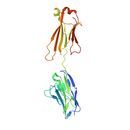
CryoEM structure of a therapeutic antibody (favezelimab) bound to human LAG3 determined using a bivalent Fab as fiducial marker.

Mishra, A.K.Shahid, S.Karade, S.S.Agnihotri, P.Kolesnikov, A.Hasan, S.S.Mariuzza, R.A.

(2023) Structure 31: 1149

  • DOI: https://doi.org/10.1016/j.str.2023.07.013
  • Primary Citation Related Structures: 
    6WKM, 8FWH, 8SO3, 8SR0

  • PubMed Abstract: 

    Lymphocyte activation gene 3 protein (LAG3) is an inhibitory receptor that is upregulated on exhausted T cells in tumors. LAG3 is a major target for cancer immunotherapy with many anti-LAG3 antibodies in clinical trials. However, there is no structural information on the epitopes recognized by these antibodies. We determined the single-particle cryoEM structure of a therapeutic antibody (favezelimab) bound to LAG3 to 3.5 Å resolution, revealing that favezelimab targets the LAG3-binding site for MHC class II, its canonical ligand. The small size of the complex between the conventional (monovalent) Fab of favezelimab and LAG3 (∼100 kDa) presented a challenge for cryoEM. Accordingly, we engineered a bivalent version of Fab favezelimab that doubled the size of the Fab-LAG3 complex and conferred a highly identifiable shape to the complex that facilitated particle selection and orientation for image processing. This study establishes bivalent Fabs as new fiducial markers for cryoEM analysis of small proteins.


  • Organizational Affiliation
    • W.M. Keck Laboratory for Structural Biology, University of Maryland Institute for Bioscience and Biotechnology Research, Rockville, MD 20850, USA; Department of Cell Biology and Molecular Genetics, University of Maryland, College Park, MD 20742, USA.

Macromolecule Content 

  • Total Structure Weight: 47.71 kDa 
  • Atom Count: 3,183 
  • Modeled Residue Count: 429 
  • Deposited Residue Count: 441 
  • Unique protein chains: 2

Macromolecules

Find similar proteins by:|  3D Structure
Entity ID: 1
MoleculeChains  Sequence LengthOrganismDetailsImage
Anti-human LAG3 (22D2) heavy chainA [auth HHH]223Mus musculusMutation(s): 0 
Find similar proteins by:|  3D Structure
Entity ID: 2
MoleculeChains  Sequence LengthOrganismDetailsImage
Anti-human LAG3 (22D2) light chainB [auth LLL]218Mus musculusMutation(s): 0 

Small Molecules

Ligands 1 Unique
IDChains Name / Formula / InChI Key2D Diagram3D Interactions
EDO
(Subject of Investigation/LOI)

Query on EDO


Download:Ideal Coordinates CCD File
C [auth LLL]1,2-ETHANEDIOL
C2 H6 O2
LYCAIKOWRPUZTN-UHFFFAOYSA-N

Experimental Data & Validation

Experimental Data

  • Method: X-RAY DIFFRACTION
  • Resolution: 2.83 Å
  • R-Value Free:  0.277 (Depositor), 0.273 (DCC) 
  • R-Value Work:  0.216 (Depositor), 0.217 (DCC) 
Space Group: C 1 2 1
Unit Cell:
Length ( Å )Angle ( ˚ )
a = 159.287α = 90
b = 44.39β = 106.136
c = 89.084γ = 90
Software Package:
Software NamePurpose
REFMACrefinement
HKL-2000data reduction
HKL-2000data scaling
PHASERphasing

Structure Validation

View Full Validation Report


Ligand Structure Quality Assessment 


Entry History 

& Funding Information

Deposition Data


Funding OrganizationLocationGrant Number
National Institutes of Health/National Institute Of Allergy and Infectious Diseases (NIH/NIAID)United StatesAI144422

Revision History  (Full details and data files)

  • Version 1.0: 2023-09-06
    Type: Initial release
  • Version 1.1: 2023-10-18
    Changes: Database references
  • Version 1.2: 2024-11-20
    Changes: Structure summary