The catalytic activity and structure of the lipid metabolizing CYP124 cytochrome P450 enzyme from Mycobacterium marinum.
Ghith, A., Bruning, J.B., Bell, S.G.(2023) Arch Biochem Biophys 737: 109554-109554
- PubMed: 36842492 Search on PubMed
- DOI: https://doi.org/10.1016/j.abb.2023.109554
- Primary Citation Related Structures:
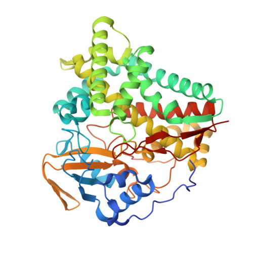
8FJO, 8FKB, 8FLO - PubMed Abstract:
The CYP124 family of cytochrome P450 enzymes, as exemplified by CYP124A1 from Mycobacterium tuberculosis, is involved in the metabolism of methyl branched lipids and cholesterol derivatives. The equivalent enzyme from Mycobacterium marinum was investigated to compare the degree of functional conservation between members of this CYP family from closely related bacteria. We compared substrate binding of each CYP124 enzyme using UV-vis spectroscopy and the catalytic oxidation of methyl branched lipids, terpenes and cholesterol derivatives was investigated. The CYP124 enzyme from M. tuberculosis displayed a larger shift to the ferric high-spin state on binding cholesterol derivatives compared to the equivalent enzyme from M. marinum. The biggest difference was observed with cholesteryl sulfate which induced distinct UV-vis spectra in each CYP124 enzyme. The selectivity for oxidation at the ω-carbon of a branched chain was maintained for all substrates, except cholesteryl sulfate which was not oxidized by either enzyme. The CYP124A1 enzyme from M. marinum, in combination with farnesol and farnesyl acetate, was structurally characterized by X-ray crystallography. These ligand-bound structures of the CYP124 enzyme revealed that the polar component of the substrates bound in a different manner to that of phytanic acid in the structure of CYP124A1 from M. tuberculosis. However, closer to the heme the structures were similar providing an explanation for the high selectivity of the enzyme for terminal methyl C-H bond oxidation. The work here demonstrates that there were differences in the biochemistry of the CYP124 enzymes from these closely related bacteria.
- Department of Chemistry, University of Adelaide, SA, 5005, Australia.
Organizational Affiliation: