8FAK | pdb_00008fak

DNA replication fork binding triggers structural changes in the PriA DNA helicase that regulate the PriA-PriB replication restart pathway in E. coli


Experimental Data Snapshot

  • Method: ELECTRON MICROSCOPY
  • Resolution: 3.22 Å
  • Aggregation State: PARTICLE 
  • Reconstruction Method: SINGLE PARTICLE 

wwPDB Validation 3D Report Full Report

Validation slider image for 8FAK

This is version 1.2 of the entry. See complete history

Literature

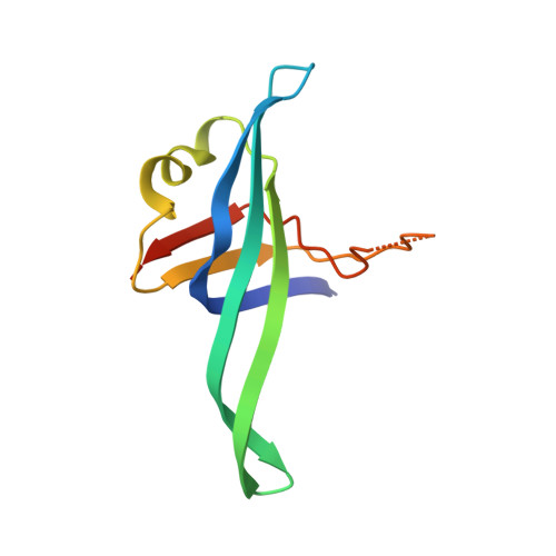
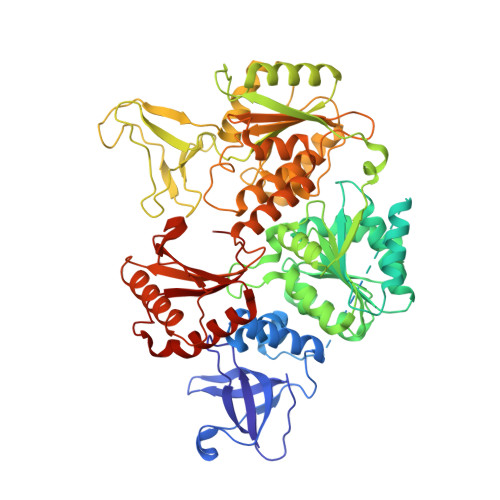
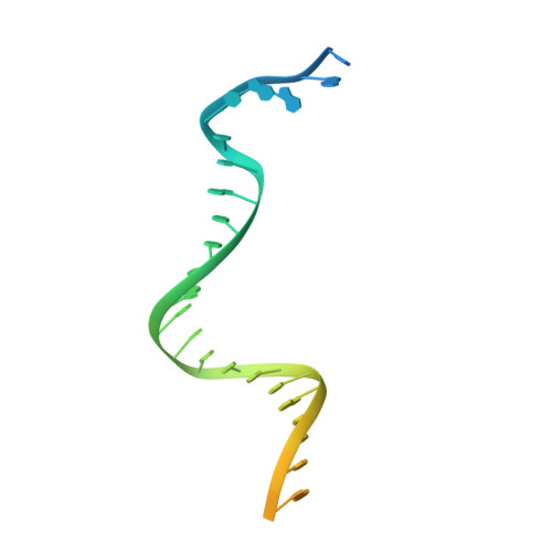
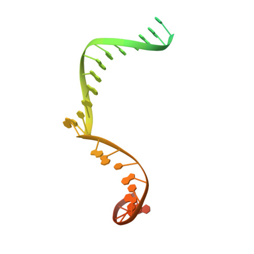
Replication fork binding triggers structural changes in the PriA helicase that govern DNA replication restart in E. coli.

Duckworth, A.T.Ducos, P.L.McMillan, S.D.Satyshur, K.A.Blumenthal, K.H.Deorio, H.R.Larson, J.A.Sandler, S.J.Grant, T.Keck, J.L.

(2023) Nat Commun 14: 2725-2725

  • DOI: https://doi.org/10.1038/s41467-023-38144-x
  • Primary Citation Related Structures: 
    8FAK

  • PubMed Abstract: 

    Bacterial replisomes often dissociate from replication forks before chromosomal replication is complete. To avoid the lethal consequences of such situations, bacteria have evolved replication restart pathways that reload replisomes onto prematurely terminated replication forks. To understand how the primary replication restart pathway in E. coli (PriA-PriB) selectively acts on replication forks, we determined the cryogenic-electron microscopy structure of a PriA/PriB/replication fork complex. Replication fork specificity arises from extensive PriA interactions with each arm of the branched DNA. These interactions reshape the PriA protein to create a pore encircling single-stranded lagging-strand DNA while also exposing a surface of PriA onto which PriB docks. Together with supporting biochemical and genetic studies, the structure reveals a switch-like mechanism for replication restart initiation in which restructuring of PriA directly couples replication fork recognition to PriA/PriB complex formation to ensure robust and high-fidelity replication re-initiation.


  • Organizational Affiliation
    • Department of Biomolecular Chemistry, University of Wisconsin-Madison, Madison, WI, 53706, USA.

Macromolecule Content 

  • Total Structure Weight: 133.99 kDa 
  • Atom Count: 7,715 
  • Modeled Residue Count: 894 
  • Deposited Residue Count: 1,035 
  • Unique protein chains: 2
  • Unique nucleic acid chains: 3

Macromolecules


Find similar proteins by:|  3D Structure
Entity ID: 1
MoleculeChains  Sequence LengthOrganismDetailsImage
Primosomal replication protein N
A, B
104Escherichia coli K-12Mutation(s): 0 
Gene Names: priBb4201JW4159
UniProt
Find proteins for P07013 (Escherichia coli (strain K12))
Explore P07013 
Go to UniProtKB:  P07013
Entity Groups
Sequence Clusters30% Identity50% Identity70% Identity90% Identity95% Identity100% Identity
UniProt GroupP07013
Sequence Annotations
Expand
Reference Sequence
Find similar proteins by:|  3D Structure
Entity ID: 3
MoleculeChains  Sequence LengthOrganismDetailsImage
Primosomal protein N'D [auth H]732Escherichia coli K-12Mutation(s): 0 
Gene Names: priAb3935JW3906
EC: 3.6.4 (PDB Primary Data), 5.6.2.4 (UniProt)
UniProt
Find proteins for P17888 (Escherichia coli (strain K12))
Explore P17888 
Go to UniProtKB:  P17888
Entity Groups
Sequence Clusters30% Identity50% Identity70% Identity90% Identity95% Identity100% Identity
UniProt GroupP17888
Sequence Annotations
Expand
Reference Sequence
Find similar nucleic acids by:  (by identity cutoff) 
Entity ID: 2
MoleculeChains LengthOrganismImage
DNA (5'-D(P*CP*AP*GP*AP*CP*TP*CP*AP*TP*TP*TP*AP*GP*CP*CP*CP*TP*TP*AP*TP*CP*CP*G)-3')C [auth G]40synthetic construct
Sequence Annotations
Expand
Reference Sequence
Find similar nucleic acids by:  (by identity cutoff) 
Entity ID: 4
MoleculeChains LengthOrganismImage
DNA (5'-D(P*CP*GP*GP*AP*TP*AP*AP*GP*GP*GP*CP*TP*GP*AP*GP*CP*AP*CP*GP*CP*CP*GP*A)-3')E [auth J]40synthetic construct
Sequence Annotations
Expand
Reference Sequence
Find similar nucleic acids by:  Sequence
Entity ID: 5
MoleculeChains LengthOrganismImage
DNA (5'-D(P*TP*CP*GP*GP*CP*GP*TP*GP*CP*TP*C)-3')F [auth Z]15synthetic construct
Sequence Annotations
Expand
Reference Sequence

Experimental Data & Validation

Experimental Data

  • Method: ELECTRON MICROSCOPY
  • Resolution: 3.22 Å
  • Aggregation State: PARTICLE 
  • Reconstruction Method: SINGLE PARTICLE 
EM Software:
TaskSoftware PackageVersion
RECONSTRUCTIONcisTEM2.0.0
MODEL REFINEMENTPHENIX

Structure Validation

View Full Validation Report


Entry History 

& Funding Information

Deposition Data


Funding OrganizationLocationGrant Number
National Institutes of Health/National Institute of General Medical Sciences (NIH/NIGMS)United StatesGM098885

Revision History  (Full details and data files)

  • Version 1.0: 2023-05-10
    Type: Initial release
  • Version 1.1: 2023-05-24
    Changes: Database references
  • Version 1.2: 2024-10-23
    Changes: Data collection, Structure summary