8F2K

Structure of yeast F1-ATPase determined with 100 micromolar cruentaren A

  • Classification: HYDROLASE
  • Organism(s): Saccharomyces cerevisiae
  • Mutation(s): No 

  • Deposited: 2022-11-08 Released: 2023-04-05 
  • Deposition Author(s): Guo, H., Rubinstein, J.L.
  • Funding Organization(s): Canada Research Chairs, Canadian Institutes of Health Research (CIHR), Canada Foundation for Innovation, National Institutes of Health/National Cancer Institute (NIH/NCI), National Institutes of Health/National Center for Advancing Translational Sciences (NIH/NCATS)

Experimental Data Snapshot

  • Method: ELECTRON MICROSCOPY
  • Resolution: 2.90 Å
  • Aggregation State: PARTICLE 
  • Reconstruction Method: SINGLE PARTICLE 

wwPDB Validation   3D Report Full Report


This is version 1.3 of the entry. See complete history


Literature

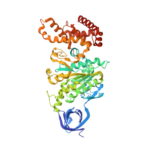
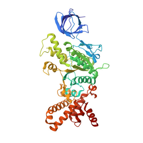
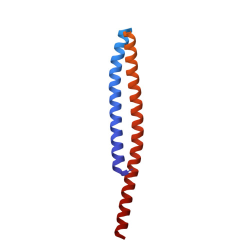
CryoEM Structure with ATP Synthase Enables Late-Stage Diversification of Cruentaren A.

Dou, X.Guo, H.D'Amico, T.Abdallah, L.Subramanian, C.Patel, B.A.Cohen, M.Rubinstein, J.L.Blagg, B.S.J.

(2023) Chemistry 29: e202300262-e202300262

  • DOI: https://doi.org/10.1002/chem.202300262
  • Primary Citation of Related Structures:  
    8F2K

  • PubMed Abstract: 

    Cruentaren A is a natural product that exhibits potent antiproliferative activity against various cancer cell lines, yet its binding site within ATP synthase remained unknown, thus limiting the development of improved analogues as anticancer agents. Herein, we report the cryogenic electron microscopy (cryoEM) structure of cruentaren A bound to ATP synthase, which allowed the design of new inhibitors through semisynthetic modification. Examples of cruentaren A derivatives include a trans-alkene isomer, which was found to exhibit similar activity to cruentaren A against three cancer cell lines as well as several other analogues that retained potent inhibitory activity. Together, these studies provide a foundation for the generation of cruentaren A derivatives as potential therapeutics for the treatment of cancer.


  • Organizational Affiliation

    Department of Chemistry and Biochemistry, The University of Notre Dame, Notre Dame, IN, 46556, USA.


Macromolecules
Find similar proteins by:  (by identity cutoff)  |  3D Structure
Entity ID: 1
MoleculeChains Sequence LengthOrganismDetailsImage
ATP synthase subunit alpha
A, B, C
485Saccharomyces cerevisiaeMutation(s): 0 
UniProt
Find proteins for P07251 (Saccharomyces cerevisiae (strain ATCC 204508 / S288c))
Explore P07251 
Go to UniProtKB:  P07251
Entity Groups  
Sequence Clusters30% Identity50% Identity70% Identity90% Identity95% Identity100% Identity
UniProt GroupP07251
Sequence Annotations
Expand
  • Reference Sequence
Find similar proteins by:  (by identity cutoff)  |  3D Structure
Entity ID: 2
MoleculeChains Sequence LengthOrganismDetailsImage
ATP synthase subunit beta
D, E, F
469Saccharomyces cerevisiaeMutation(s): 0 
EC: 7.1.2.2
UniProt
Find proteins for P00830 (Saccharomyces cerevisiae (strain ATCC 204508 / S288c))
Explore P00830 
Go to UniProtKB:  P00830
Entity Groups  
Sequence Clusters30% Identity50% Identity70% Identity90% Identity95% Identity100% Identity
UniProt GroupP00830
Sequence Annotations
Expand
  • Reference Sequence
Find similar proteins by:  (by identity cutoff)  |  3D Structure
Entity ID: 3
MoleculeChains Sequence LengthOrganismDetailsImage
ATP synthase subunit gamma275Saccharomyces cerevisiaeMutation(s): 0 
UniProt
Find proteins for P38077 (Saccharomyces cerevisiae (strain ATCC 204508 / S288c))
Explore P38077 
Go to UniProtKB:  P38077
Entity Groups  
Sequence Clusters30% Identity50% Identity70% Identity90% Identity95% Identity100% Identity
UniProt GroupP38077
Sequence Annotations
Expand
  • Reference Sequence
Small Molecules
Ligands 3 Unique
IDChains Name / Formula / InChI Key2D Diagram3D Interactions
XBC (Subject of Investigation/LOI)
Query on XBC

Download Ideal Coordinates CCD File 
P [auth E],
S [auth F]
Cruentaren A
C33 H51 N O8
UXPPKIOUXFFKDI-QGMVIAOPSA-N
ATP
Query on ATP

Download Ideal Coordinates CCD File 
H [auth A],
J [auth B],
L [auth C],
N [auth E],
Q [auth F]
ADENOSINE-5'-TRIPHOSPHATE
C10 H16 N5 O13 P3
ZKHQWZAMYRWXGA-KQYNXXCUSA-N
MG
Query on MG

Download Ideal Coordinates CCD File 
I [auth A],
K [auth B],
M [auth C],
O [auth E],
R [auth F]
MAGNESIUM ION
Mg
JLVVSXFLKOJNIY-UHFFFAOYSA-N
Experimental Data & Validation

Experimental Data

  • Method: ELECTRON MICROSCOPY
  • Resolution: 2.90 Å
  • Aggregation State: PARTICLE 
  • Reconstruction Method: SINGLE PARTICLE 
EM Software:
TaskSoftware PackageVersion
MODEL REFINEMENTCoot0.9.8
MODEL REFINEMENTISOLDE1.4.0
MODEL REFINEMENTPHENIX1.20.1
RECONSTRUCTIONcryoSPARC3.1.0

Structure Validation

View Full Validation Report


Entry History & Funding Information

Deposition Data


Funding OrganizationLocationGrant Number
Canada Research ChairsCanadaElectron Cryomicroscopy
Canadian Institutes of Health Research (CIHR)CanadaPJT162186
Canada Foundation for InnovationCanada--
National Institutes of Health/National Cancer Institute (NIH/NCI)United StatesR01 CA216919
National Institutes of Health/National Center for Advancing Translational Sciences (NIH/NCATS)United StatesUL1TR002529

Revision History  (Full details and data files)

  • Version 1.0: 2023-04-05
    Type: Initial release
  • Version 1.1: 2023-10-18
    Changes: Data collection, Database references, Refinement description
  • Version 1.2: 2024-04-17
    Changes: Database references
  • Version 1.3: 2024-04-24
    Changes: Database references