8F0P | pdb_00008f0p

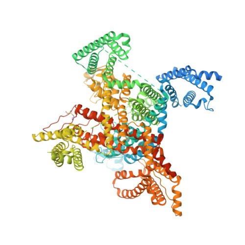
Structure of VSD4-NaV1.7-NaVPas channel chimera bound to the hybrid inhibitor GNE-1305


Experimental Data Snapshot

  • Method: ELECTRON MICROSCOPY
  • Resolution: 2.20 Å
  • Aggregation State: PARTICLE 
  • Reconstruction Method: SINGLE PARTICLE 

wwPDB Validation 3D Report Full Report

Validation slider image for 8F0P

This is version 1.0 of the entry. See complete history. 

Literature

Cryo-EM reveals an unprecedented binding site for Na V 1.7 inhibitors enabling rational design of potent hybrid inhibitors.

Kschonsak, M., Jao, C.C., Arthur, C.P., Rohou, A.L., Bergeron, P., Ortwine, D.F., McKerrall, S.J., Hackos, D.H., Deng, L., Chen, J., Li, T., Dragovich, P.S., Volgraf, M., Wright, M.R., Payandeh, J., Ciferri, C., Tellis, J.C.

(2023) Elife 12

  • DOI: https://doi.org/10.7554/eLife.84151
  • Primary Citation Related Structures: 
    8F0P, 8F0Q, 8F0R, 8F0S

  • PubMed Abstract: 

    The voltage-gated sodium (Na V ) channel Na V 1.7 has been identified as a potential novel analgesic target due to its involvement in human pain syndromes. However, clinically available Na V channel-blocking drugs are not selective among the nine Na V channel subtypes, Na V 1.1-Na V 1.9. Moreover, the two currently known classes of Na V 1.7 subtype-selective inhibitors (aryl- and acylsulfonamides) have undesirable characteristics that may limit their development. To this point understanding of the structure-activity relationships of the acylsulfonamide class of Na V 1.7 inhibitors, exemplified by the clinical development candidate GDC-0310 , has been based solely on a single co-crystal structure of an arylsulfonamide inhibitor bound to voltage-sensing domain 4 (VSD4). To advance inhibitor design targeting the Na V 1.7 channel, we pursued high-resolution ligand-bound Na V 1.7-VSD4 structures using cryogenic electron microscopy (cryo-EM). Here, we report that GDC-0310 engages the Na V 1.7-VSD4 through an unexpected binding mode orthogonal to the arylsulfonamide inhibitor class binding pose, which identifies a previously unknown ligand binding site in Na V channels. This finding enabled the design of a novel hybrid inhibitor series that bridges the aryl- and acylsulfonamide binding pockets and allows for the generation of molecules with substantially differentiated structures and properties. Overall, our study highlights the power of cryo-EM methods to pursue challenging drug targets using iterative and high-resolution structure-guided inhibitor design. This work also underscores an important role of the membrane bilayer in the optimization of selective Na V channel modulators targeting VSD4.


  • Organizational Affiliation: 
    • Genentech Inc, Structural Biology, South San Francisco, United States.

Macromolecule Content 

  • Total Structure Weight: 190.81 kDa 
  • Atom Count: 9,616 
  • Modeled Residue Count: 1,116 
  • Deposited Residue Count: 1,608 
  • Unique protein chains: 1

Macromolecules

Find similar proteins by:|  3D Structure
Entity ID: 1
MoleculeChains  Sequence LengthOrganismDetailsImage
Sodium channel protein PaFPC1,Sodium channel protein type 9 subunit alpha chimera1,608Homo sapiens, Periplaneta americanaMutation(s): 0 
Membrane Entity: Yes 
UniProt & NIH Common Fund Data Resources
Find proteins for D0E0C2 (Periplaneta americana)
Explore D0E0C2 
Go to UniProtKB:  D0E0C2
Find proteins for Q15858 (Homo sapiens)
Explore Q15858 
Go to UniProtKB:  Q15858
PHAROS:  Q15858
GTEx:  ENSG00000169432 
Entity Groups
Sequence Clusters30% Identity50% Identity70% Identity90% Identity95% Identity100% Identity
UniProt GroupsQ15858D0E0C2
Glycosylation
Glycosylation Sites: 4Go to GlyGen: Q15858-1
Sequence Annotations
Expand
Reference Sequence

Oligosaccharides

Help  
Entity ID: 2
MoleculeChains Length2D Diagram GlycosylationD Interactions
beta-D-mannopyranose-(1-3)-[beta-D-mannopyranose-(1-6)]beta-D-mannopyranose-(1-3)-2-acetamido-2-deoxy-beta-D-glucopyranose-(1-3)-2-acetamido-2-deoxy-beta-D-glucopyranose
B
5N/AN-Glycosylation
Glycosylation Resources
GlyTouCan: G61217QH
GlyCosmos: G61217QH
GlyGen: G61217QH

Small Molecules

Ligands 4 Unique
IDChains Name / Formula / InChI Key2D Diagram3D Interactions
PEE

Query on PEE


Download:Ideal Coordinates CCD File
F [auth A],
G [auth A],
H [auth A],
I [auth A],
J [auth A]
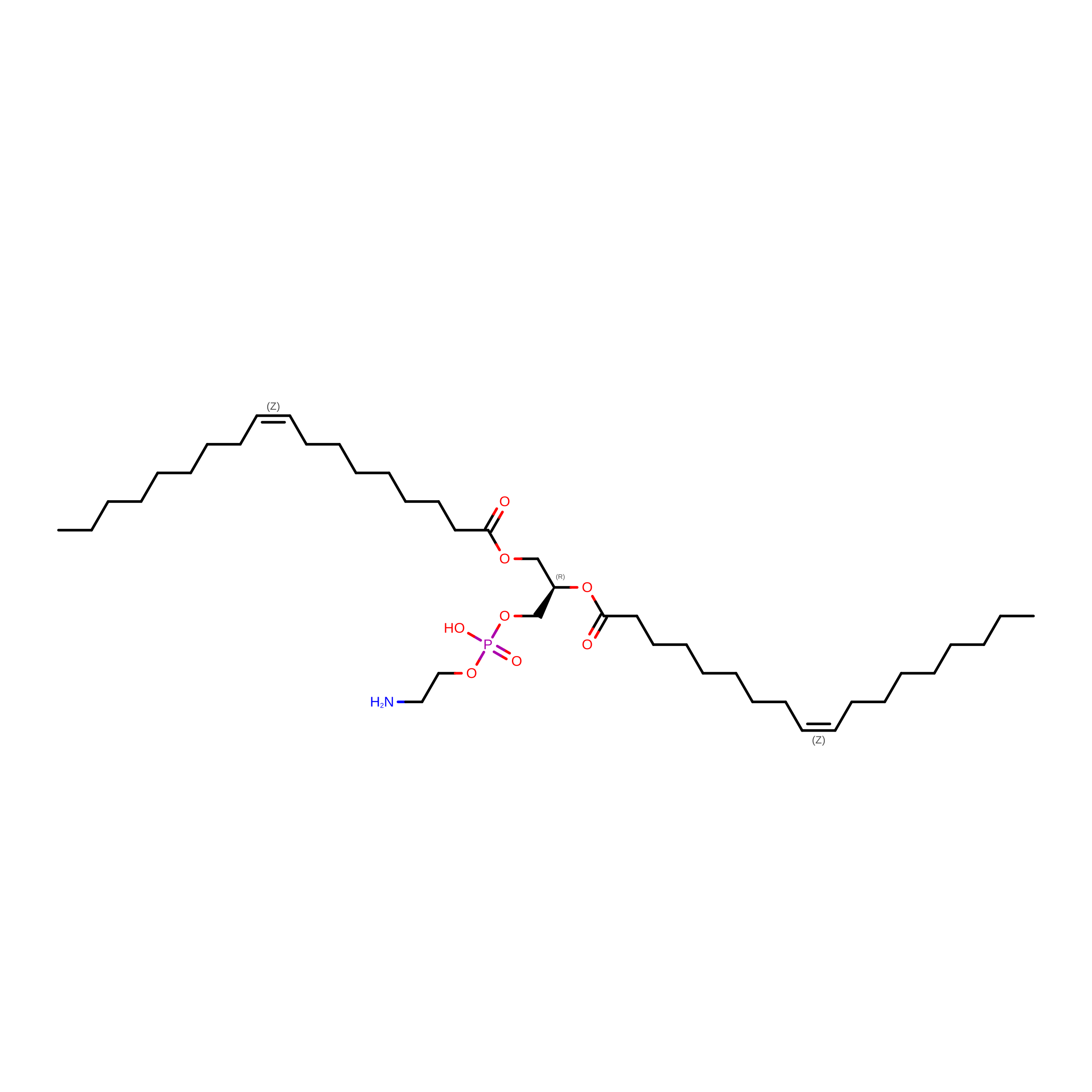
1,2-dioleoyl-sn-glycero-3-phosphoethanolamine
C41 H78 N O8 P
MWRBNPKJOOWZPW-NYVOMTAGSA-N
X7L
(Subject of Investigation/LOI)

Query on X7L


Download:Ideal Coordinates CCD File
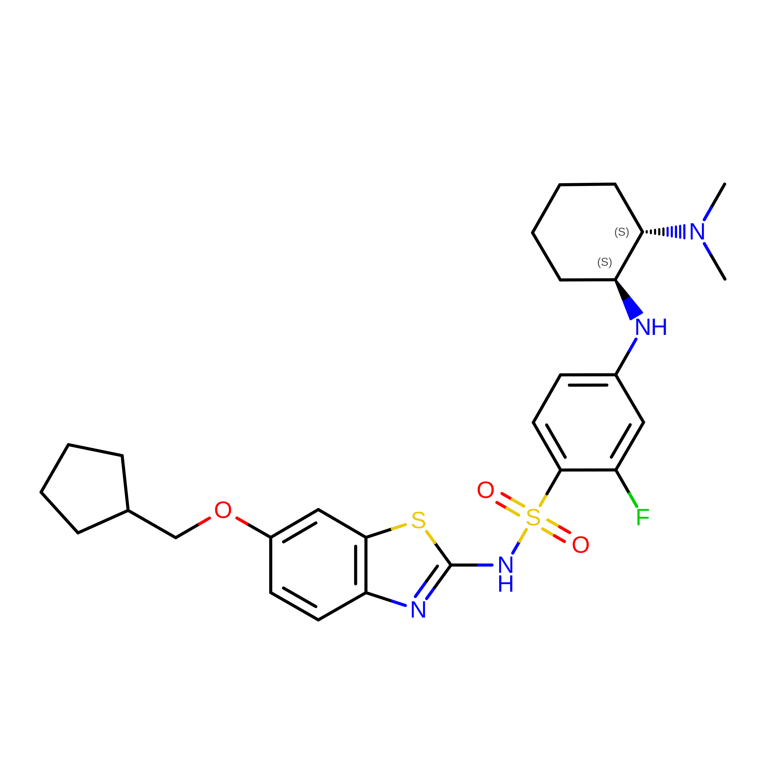
L [auth A]N-[6-(cyclopentylmethoxy)-1,3-benzothiazol-2-yl]-4-{[(1S,2S)-2-(dimethylamino)cyclohexyl]amino}-2-fluorobenzene-1-sulfonamide
C27 H35 F N4 O3 S2
DYROPBGPVFEZHO-UPVQGACJSA-N
Y01

Query on Y01


Download:Ideal Coordinates CCD File
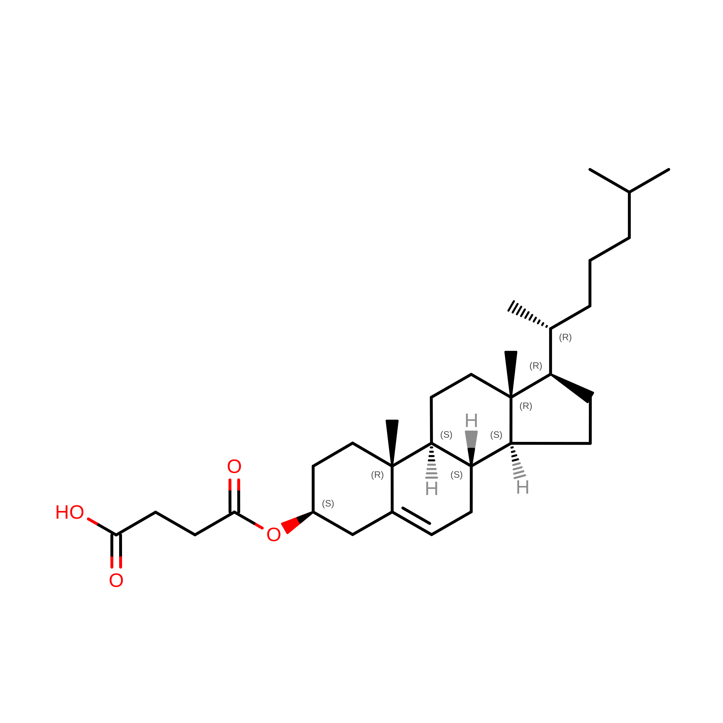
K [auth A]CHOLESTEROL HEMISUCCINATE
C31 H50 O4
WLNARFZDISHUGS-MIXBDBMTSA-N
NAG

Query on NAG


Download:Ideal Coordinates CCD File
C [auth A],
D [auth A],
E [auth A]
2-acetamido-2-deoxy-beta-D-glucopyranose
C8 H15 N O6
OVRNDRQMDRJTHS-FMDGEEDCSA-N

Experimental Data & Validation

Experimental Data

  • Method: ELECTRON MICROSCOPY
  • Resolution: 2.20 Å
  • Aggregation State: PARTICLE 
  • Reconstruction Method: SINGLE PARTICLE 
EM Software:
TaskSoftware PackageVersion
MODEL REFINEMENTPHENIX1.20
RECONSTRUCTIONPHENIX1.20

Structure Validation

View Full Validation Report


Entry History 

& Funding Information

Deposition Data


Funding OrganizationLocationGrant Number
Not funded--

Revision History  (Full details and data files)

  • Version 1.0: 2023-04-12
    Type: Initial release