Structural and Mechanistic Basis for the Inactivation of Human Ornithine Aminotransferase by (3 S ,4 S )-3-Amino-4-fluorocyclopentenecarboxylic Acid.
Shen, S., Butrin, A., Beaupre, B.A., Ferreira, G.M., Doubleday, P.F., Grass, D.H., Zhu, W., Kelleher, N.L., Moran, G.R., Liu, D., Silverman, R.B.(2023) Molecules 28
- PubMed: 36770800 Search on PubMedSearch on PubMed Central
- DOI: https://doi.org/10.3390/molecules28031133
- Primary Citation Related Structures:
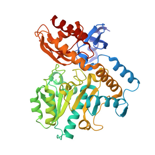
8EZ1 - PubMed Abstract:
Ornithine aminotransferase (OAT) is overexpressed in hepatocellular carcinoma (HCC), and we previously showed that inactivation of OAT inhibits the growth of HCC. Recently, we found that (3 S ,4 S )-3-amino-4-fluorocyclopentenecarboxylic acid ( 5 ) was a potent inactivator of γ-aminobutyric acid aminotransferase (GABA-AT), proceeding by an enamine mechanism. Here we describe our investigations into the activity and mechanism of 5 as an inactivator of human OAT. We have found that 5 exhibits 10-fold less inactivation efficiency ( k inact / K I ) against h OAT than GABA-AT. A comprehensive mechanistic study was carried out to understand its inactivation mechanism with h OAT. p K a and electrostatic potential calculations were performed to further support the notion that the α,β-unsaturated alkene of 5 is critical for enhancing acidity and nucleophilicity of the corresponding intermediates and ultimately responsible for the improved inactivation efficiency of 5 over the corresponding saturated analogue ( 4 ). Intact protein mass spectrometry and the crystal structure complex with h OAT provide evidence to conclude that 5 mainly inactivates h OAT through noncovalent interactions, and that, unlike with GABA-AT, covalent binding with h OAT is a minor component of the total inhibition which is unique relative to other monofluoro-substituted derivatives. Furthermore, based on the results of transient-state measurements and free energy calculations, it is suggested that the α,β-unsaturated carboxylate group of PLP-bound 5 may be directly involved in the inactivation cascade by forming an enolate intermediate. Overall, compound 5 exhibits unusual structural conversions which are catalyzed by specific residues within h OAT, ultimately leading to an enamine mechanism-based inactivation of h OAT through noncovalent interactions and covalent modification.
- Department of Chemistry and Center for Developmental Therapeutics, Northwestern University, Evanston, IL 60208, USA.
Organizational Affiliation: