8EOH

crystal structure of human Cytochrome P450 8B1 in complex with a C12-Pyridine Containing Steroid

  • Classification: OXIDOREDUCTASE
  • Organism(s): Homo sapiens
  • Expression System: Escherichia coli
  • Mutation(s): No 

  • Deposited: 2022-10-03 Released: 2023-08-02 
  • Deposition Author(s): Liu, J., Scott, E.E.
  • Funding Organization(s): National Institutes of Health/National Institute of General Medical Sciences (NIH/NIGMS)

Experimental Data Snapshot

  • Method: X-RAY DIFFRACTION
  • Resolution: 2.65 Å
  • R-Value Free: 0.265 
  • R-Value Work: 0.223 
  • R-Value Observed: 0.225 

wwPDB Validation   3D Report Full Report


Ligand Structure Quality Assessment 


This is version 1.0 of the entry. See complete history


Literature

Pyridine-containing substrate analogs are restricted from accessing the human cytochrome P450 8B1 active site by tryptophan 281.

Liu, J.Offei, S.D.Yoshimoto, F.K.Scott, E.E.

(2023) J Biol Chem 299: 103032-103032

  • DOI: https://doi.org/10.1016/j.jbc.2023.103032
  • Primary Citation of Related Structures:  
    8EOH

  • PubMed Abstract: 

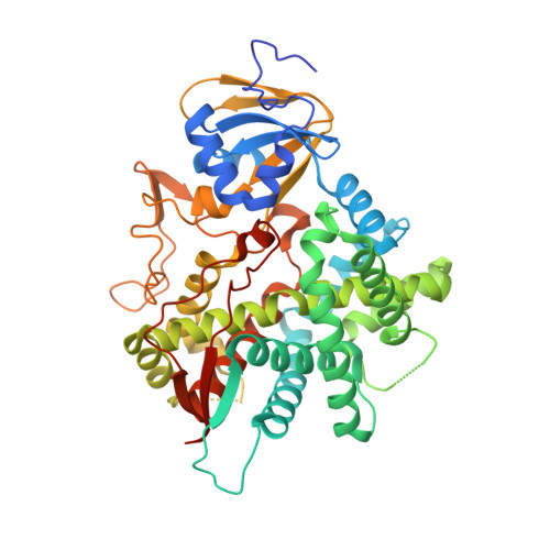
    The human oxysterol 12α-hydroxylase cytochrome P450 8B1 (CYP8B1) is a validated drug target for both type 2 diabetes and nonalcoholic fatty liver disease, but effective selective inhibitors are not yet available. Herein, steroidal substrate-mimicking compounds with a pyridine ring appended to the C12 site of metabolism were designed as inhibitors, synthesized, and evaluated in terms of their functional and structural interactions with CYP8B1. While the pyridine nitrogen was intended to coordinate the CYP8B1 active site heme iron, none of these compounds elicited shifts in the CYP8B1 Soret absorbance consistent with this type of interaction. However, when CYP8B1 was cocrystallized with the pyridine-containing compound with the 3-keto-Δ4 steroid backbone most similar to the endogenous substrate, it was apparent that this ligand was bound in a channel leading to the active site, instead of near the heme iron. Inspection of this structure suggested that tryptophan 281 directly above the heme might restrict active site binding of potential inhibitors with this design. This hypothesis was supported when a CYP8B1 W281F mutation did allow all three compounds to coordinate the heme iron as designed. These results indicated that the design of next-generation CYP8B1 inhibitors should be compatible with the low-ceiling tryptophan immediately above the heme iron.


  • Organizational Affiliation

    Department of Medicinal Chemistry, University of Michigan, Ann Arbor, Michigan, USA.


Macromolecules
Find similar proteins by:  (by identity cutoff)  |  3D Structure
Entity ID: 1
MoleculeChains Sequence LengthOrganismDetailsImage
7-alpha-hydroxycholest-4-en-3-one 12-alpha-hydroxylase489Homo sapiensMutation(s): 0 
Gene Names: CYP8B1CYP12
EC: 1.14.18.8
UniProt & NIH Common Fund Data Resources
Find proteins for Q9UNU6 (Homo sapiens)
Explore Q9UNU6 
Go to UniProtKB:  Q9UNU6
PHAROS:  Q9UNU6
GTEx:  ENSG00000180432 
Entity Groups  
Sequence Clusters30% Identity50% Identity70% Identity90% Identity95% Identity100% Identity
UniProt GroupQ9UNU6
Sequence Annotations
Expand
  • Reference Sequence
Small Molecules
Ligands 2 Unique
IDChains Name / Formula / InChI Key2D Diagram3D Interactions
HEM (Subject of Investigation/LOI)
Query on HEM

Download Ideal Coordinates CCD File 
B [auth A]PROTOPORPHYRIN IX CONTAINING FE
C34 H32 Fe N4 O4
KABFMIBPWCXCRK-RGGAHWMASA-L
WOL (Subject of Investigation/LOI)
Query on WOL

Download Ideal Coordinates CCD File 
C [auth A]12-(pyridin-3-yl)-8alpha,10alpha,13alpha,14beta-androsta-4,11-diene-3,17-dione
C24 H27 N O2
XWNGZWLWVUCVNS-ZGJXEWGXSA-N
Experimental Data & Validation

Experimental Data

  • Method: X-RAY DIFFRACTION
  • Resolution: 2.65 Å
  • R-Value Free: 0.265 
  • R-Value Work: 0.223 
  • R-Value Observed: 0.225 
  • Space Group: P 21 21 21
Unit Cell:
Length ( Å )Angle ( ˚ )
a = 62.127α = 90
b = 90.124β = 90
c = 103.018γ = 90
Software Package:
Software NamePurpose
PHENIXrefinement
Blu-Icedata collection
SCALAdata scaling
PHASERphasing
Cootmodel building
XDSdata reduction

Structure Validation

View Full Validation Report


Ligand Structure Quality Assessment 


Entry History & Funding Information

Deposition Data


Funding OrganizationLocationGrant Number
National Institutes of Health/National Institute of General Medical Sciences (NIH/NIGMS)United States020450

Revision History  (Full details and data files)

  • Version 1.0: 2023-08-02
    Type: Initial release