8ENC

Helical reconstruction of the human cardiac actin-tropomyosin-myosin loop 4 7G mutant complex


Experimental Data Snapshot

  • Method: ELECTRON MICROSCOPY
  • Resolution: 3.60 Å
  • Aggregation State: FILAMENT 
  • Reconstruction Method: HELICAL 

wwPDB Validation   3D Report Full Report


This is version 1.1 of the entry. See complete history


Literature

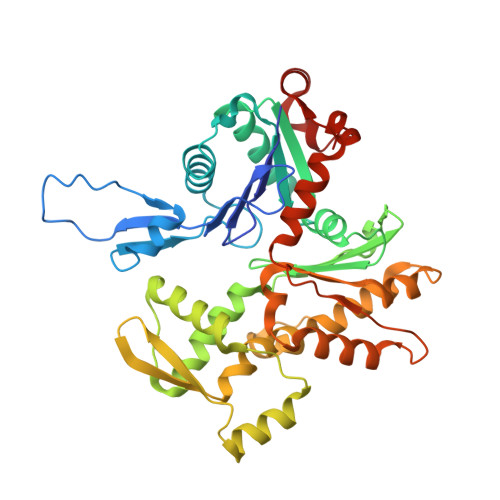
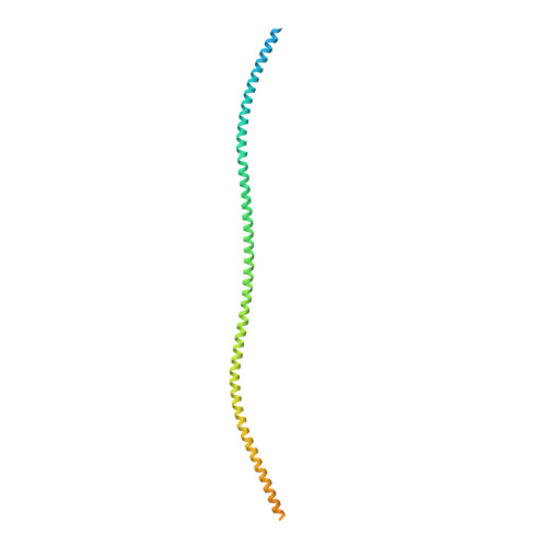
Myosin loop-4 is critical for optimal tropomyosin repositioning on actin during muscle activation and relaxation.

Doran, M.H.Rynkiewicz, M.J.Pavadai, E.Bodt, S.M.L.Rasicci, D.Moore, J.R.Yengo, C.M.Bullitt, E.Lehman, W.

(2023) J Gen Physiol 155

  • DOI: https://doi.org/10.1085/jgp.202213274
  • Primary Citation of Related Structures:  
    8EFI, 8ENC

  • PubMed Abstract: 

    During force-generating steps of the muscle crossbridge cycle, the tip of the myosin motor, specifically loop-4, contacts the tropomyosin cable of actin filaments. In the current study, we determined the corresponding effect of myosin loop-4 on the regulatory positioning of tropomyosin on actin. To accomplish this, we compared high-resolution cryo-EM structures of myosin S1-decorated thin filaments containing either wild-type or a loop-4 mutant construct, where the seven-residue portion of myosin loop-4 that contacts tropomyosin was replaced by glycine residues, thus removing polar side chains from residues 366-372. Cryo-EM analysis of fully decorated actin-tropomyosin filaments with wild-type and mutant S1, yielded 3.4-3.6 Å resolution reconstructions, with even higher definition at the actin-myosin interface. Loop-4 densities both in wild-type and mutant S1 were clearly identified, and side chains were resolved in the wild-type structure. Aside from loop-4, actin and myosin structural domains were indistinguishable from each other when filaments were decorated with either mutant or wild-type S1. In marked contrast, the position of tropomyosin on actin in the two reconstructions differed by 3 to 4 Å. In maps of filaments containing the mutant, tropomyosin was located closer to the myosin-head and thus moved in the direction of the C-state conformation adopted by myosin-free thin filaments. Complementary interaction energy measurements showed that tropomyosin in the mutant thin filaments sits on actin in a local energy minimum, whereas tropomyosin is positioned by wild-type S1 in an energetically unfavorable location. We propose that the high potential energy associated with tropomyosin positioning in wild-type filaments favors an effective transition to B- and C-states following release of myosin from the thin filaments during relaxation.


  • Organizational Affiliation

    Department of Physiology & Biophysics, Boston University Chobanian & Avedisian School of Medicine, Boston, MA.


Macromolecules
Find similar proteins by:  (by identity cutoff)  |  3D Structure
Entity ID: 1
MoleculeChains Sequence LengthOrganismDetailsImage
Myosin-7A [auth M]1,935Homo sapiensMutation(s): 7 
Gene Names: MYH7MYHCB
UniProt & NIH Common Fund Data Resources
Find proteins for P12883 (Homo sapiens)
Explore P12883 
Go to UniProtKB:  P12883
PHAROS:  P12883
GTEx:  ENSG00000092054 
Entity Groups  
Sequence Clusters30% Identity50% Identity70% Identity90% Identity95% Identity100% Identity
UniProt GroupP12883
Sequence Annotations
Expand
  • Reference Sequence
Find similar proteins by:  (by identity cutoff)  |  3D Structure
Entity ID: 2
MoleculeChains Sequence LengthOrganismDetailsImage
Actin, alpha cardiac muscle 1
B, C, D, E, F
377Sus scrofaMutation(s): 0 
UniProt
Find proteins for B6VNT8 (Sus scrofa)
Explore B6VNT8 
Go to UniProtKB:  B6VNT8
Entity Groups  
Sequence Clusters30% Identity50% Identity70% Identity90% Identity95% Identity100% Identity
UniProt GroupB6VNT8
Sequence Annotations
Expand
  • Reference Sequence
Find similar proteins by:  (by identity cutoff)  |  3D Structure
Entity ID: 3
MoleculeChains Sequence LengthOrganismDetailsImage
Tropomyosin alpha-1 chainG [auth O],
H [auth P]
284Homo sapiensMutation(s): 0 
Gene Names: TPM1C15orf13TMSA
UniProt & NIH Common Fund Data Resources
Find proteins for P09493 (Homo sapiens)
Explore P09493 
Go to UniProtKB:  P09493
PHAROS:  P09493
GTEx:  ENSG00000140416 
Entity Groups  
Sequence Clusters30% Identity50% Identity70% Identity90% Identity95% Identity100% Identity
UniProt GroupP09493
Sequence Annotations
Expand
  • Reference Sequence
Small Molecules
Ligands 2 Unique
IDChains Name / Formula / InChI Key2D Diagram3D Interactions
ADP
Query on ADP

Download Ideal Coordinates CCD File 
I [auth B],
K [auth C],
M [auth D],
O [auth E],
Q [auth F]
ADENOSINE-5'-DIPHOSPHATE
C10 H15 N5 O10 P2
XTWYTFMLZFPYCI-KQYNXXCUSA-N
MG
Query on MG

Download Ideal Coordinates CCD File 
J [auth B],
L [auth C],
N [auth D],
P [auth E],
R [auth F]
MAGNESIUM ION
Mg
JLVVSXFLKOJNIY-UHFFFAOYSA-N
Experimental Data & Validation

Experimental Data

  • Method: ELECTRON MICROSCOPY
  • Resolution: 3.60 Å
  • Aggregation State: FILAMENT 
  • Reconstruction Method: HELICAL 
EM Software:
TaskSoftware PackageVersion
MODEL REFINEMENTPHENIX1.18
RECONSTRUCTIONRELION

Structure Validation

View Full Validation Report


Entry History & Funding Information

Deposition Data


Funding OrganizationLocationGrant Number
National Institutes of Health/National Heart, Lung, and Blood Institute (NIH/NHLBI)United StatesR01HL036153

Revision History  (Full details and data files)

  • Version 1.0: 2022-11-23
    Type: Initial release
  • Version 1.1: 2022-12-14
    Changes: Database references, Refinement description