Computational design of potent and selective binders of BAK and BAX.
Berger, S., Lee, E.F., Harris, T.J., Tran, S., Bera, A.K., Arguinchona, L., Kang, A., Sankaran, B., Kasapgil, S., Miller, M.S., Smyth, S., Lutfi, M., Uren, R.T., Kluck, R.M., Colman, P.M., Fairlie, W.D., Czabotar, P.E., Baker, D., Birkinshaw, R.W.(2025) Sci Adv 11: eadt4170-eadt4170
- PubMed: 40911686 Search on PubMedSearch on PubMed Central
- DOI: https://doi.org/10.1126/sciadv.adt4170
- Primary Citation Related Structures:
8EJA, 9CLB - PubMed Abstract:
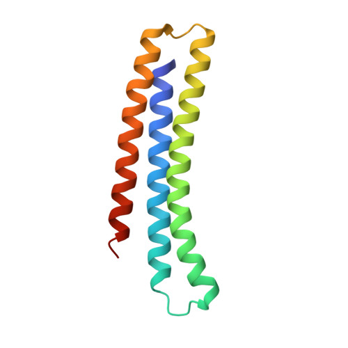
Potent and selective binders of the key proapoptotic proteins BAK and BAX have not been described. We use computational protein design to generate high affinity binders of BAK and BAX with greater than 100-fold specificity for their target. Both binders activate their targets when at low concentration, driving pore formation, but inhibit membrane permeabilization when in excess. Crystallography shows that the BAK binder induces BAK unfolding, exposing the α6 helix and BH3 domain. Together, these data suggest that upon binding, BAK or BAX unfold; at high binder concentrations, self-association of the partially folded BAK or BAX proteins is blocked and the membrane remains intact, whereas at low concentrations, dimers form, and the membrane ruptures. Our designed binders modulate apoptosis via direct, specific interactions with BAK and BAX and reveal that for therapeutic strategies targeting BAK and BAX, inhibition requires saturating binder concentrations at the site of action.
- Department of Biochemistry, University of Washington, Seattle, WA 98195, USA.
Organizational Affiliation: