Human and bacterial TatD enzymes exhibit apurinic/apyrimidinic (AP) endonuclease activity.
Dorival, J., Eichman, B.F.(2023) Nucleic Acids Res 51: 2838-2849
- PubMed: 36881763 Search on PubMedSearch on PubMed Central
- DOI: https://doi.org/10.1093/nar/gkad133
- Primary Citation Related Structures:
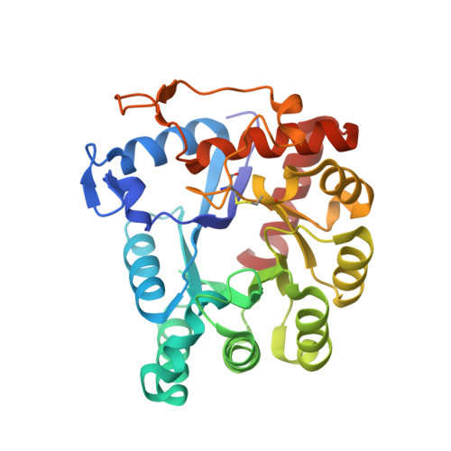
8EFG - PubMed Abstract:
TatD enzymes are evolutionarily conserved deoxyribonucleases associated with DNA repair, apoptosis, development, and parasite virulence. Three TatD paralogs exist in humans, but their nuclease functions are unknown. Here, we describe the nuclease activities of two of the three human TatD paralogs, TATDN1 and TATDN3, which represent two phylogenetically distinct clades based on unique active site motifs. We found that in addition to 3'-5' exonuclease activity associated with other TatD proteins, both TATDN1 and TATDN3 exhibited apurinic/apyrimidinic (AP) endonuclease activity. The AP endonuclease activity was observed only in double-stranded DNA, whereas exonuclease activity was operative primarily in single-stranded DNA. Both nuclease activities were observed in the presence of Mg2+ or Mn2+, and we identified several divalent metal cofactors that inhibited exonuclease and supported AP endonuclease activity. Biochemical analysis and a crystal structure of TATDN1 bound to 2'-deoxyadenosine 5'-monophosphate in the active site are consistent with two-metal ion catalysis, and we identify several residues that differentiate nuclease activities in the two proteins. In addition, we show that the three Escherichia coli TatD paralogs are also AP endonucleases, indicating that this activity is conserved across evolution. Together, these results indicate that TatD enzymes constitute a family of ancient AP endonucleases.
- Department of Biological Sciences, Vanderbilt University, Nashville, TN, USA.
Organizational Affiliation: