


Structure of Ternary Complex of cGAS with dsDNA and Bound 5-pppI(2,5)pA
Wu, S., Sohn, J.To be published.
Experimental Data Snapshot
Starting Model: experimental
View more details
Entity ID: 1 | |||||
|---|---|---|---|---|---|
| Molecule | Chains | Sequence Length | Organism | Details | Image |
| Cyclic GMP-AMP synthase | A, B [auth C] | 364 | Mus musculus | Mutation(s): 0 EC: 2.7.7.86 | 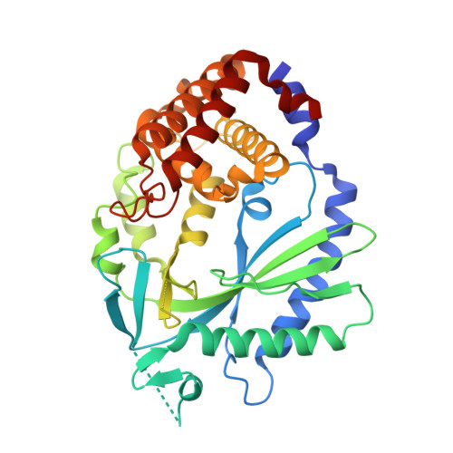 |
UniProt & NIH Common Fund Data Resources | |||||
IMPC: MGI:2442261 | |||||
Entity Groups | |||||
| Sequence Clusters | 30% Identity50% Identity70% Identity90% Identity95% Identity100% Identity | ||||
| UniProt Group | Q8C6L5 | ||||
Sequence AnnotationsExpand | |||||
Reference Sequence | |||||
Entity ID: 2 | ||||
| Molecule | Chains | Length | Organism | Image |
|---|---|---|---|---|
| Palindromic DNA18 | C [auth E], D [auth F], E [auth I], F [auth J] | 18 | synthetic construct | 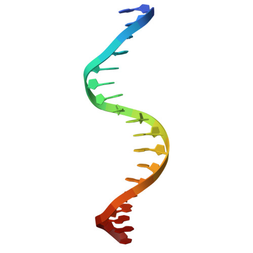 |
Sequence AnnotationsExpand | ||||
Reference Sequence | ||||
| Ligands 4 Unique | |||||
|---|---|---|---|---|---|
| ID | Chains | Name / Formula / InChI Key | 2D Diagram | 3D Interactions | |
| VWX (Subject of Investigation/LOI) Download:Ideal Coordinates CCD File | J [auth C] | [[(2~{R},3~{R},4~{R},5~{R})-4-[[(2~{R},3~{S},4~{R},5~{R})-5-(6-aminopurin-9-yl)-3,4-bis(oxidanyl)oxolan-2-yl]methoxy-oxidanyl-phosphoryl]oxy-3-oxidanyl-5-(6-oxidanylidene-1~{H}-purin-9-yl)oxolan-2-yl]methoxy-oxidanyl-phosphoryl] phosphono hydrogen phosphate C20 H27 N9 O20 P4 SWVKSPSEHJCWAI-XPWFQUROSA-N |  | ||
| ATP Download:Ideal Coordinates CCD File | G [auth A] | ADENOSINE-5'-TRIPHOSPHATE C10 H16 N5 O13 P3 ZKHQWZAMYRWXGA-KQYNXXCUSA-N |  | ||
| ZN Download:Ideal Coordinates CCD File | I [auth A], L [auth C] | ZINC ION Zn PTFCDOFLOPIGGS-UHFFFAOYSA-N |  | ||
| MG Download:Ideal Coordinates CCD File | H [auth A], K [auth C] | MAGNESIUM ION Mg JLVVSXFLKOJNIY-UHFFFAOYSA-N |  | ||
| Length ( Å ) | Angle ( ˚ ) |
|---|---|
| a = 77.045 | α = 90 |
| b = 98.534 | β = 90 |
| c = 141.706 | γ = 90 |
| Software Name | Purpose |
|---|---|
| XDS | data reduction |
| Aimless | data scaling |
| MOLREP | phasing |
| Coot | model building |
| REFMAC | refinement |
| PDB_EXTRACT | data extraction |
| Funding Organization | Location | Grant Number |
|---|---|---|
| National Institutes of Health/National Institute of General Medical Sciences (NIH/NIGMS) | United States | -- |


