8E0R

Crystal structure of mouse APCDD1 in P21 space group


Experimental Data Snapshot

  • Method: X-RAY DIFFRACTION
  • Resolution: 1.95 Å
  • R-Value Free: 0.247 
  • R-Value Work: 0.209 
  • R-Value Observed: 0.211 

wwPDB Validation   3D Report Full Report


This is version 1.2 of the entry. See complete history


Literature

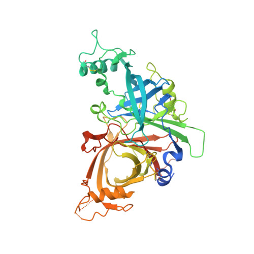
Structure of WNT inhibitor adenomatosis polyposis coli down-regulated 1 (APCDD1), a cell-surface lipid-binding protein.

Hsieh, F.L.Chang, T.H.Gabelli, S.B.Nathans, J.

(2023) Proc Natl Acad Sci U S A 120: e2217096120-e2217096120

  • DOI: https://doi.org/10.1073/pnas.2217096120
  • Primary Citation of Related Structures:  
    8E0P, 8E0R, 8E0W

  • PubMed Abstract: 

    Diverse extracellular proteins negatively regulate WNT signaling. One such regulator is adenomatosis polyposis coli down-regulated 1 (APCDD1), a conserved single-span transmembrane protein. In response to WNT signaling in a variety of tissues, APCDD1 transcripts are highly up-regulated. We have determined the three-dimensional structure of the extracellular domain of APCDD1, and this structure reveals an unusual architecture consisting of two closely apposed β-barrel domains (ABD1 and ABD2). ABD2, but not ABD1, has a large hydrophobic pocket that accommodates a bound lipid. The APCDD1 ECD can also bind to WNT7A, presumably via its covalently bound palmitoleate, a modification that is common to all WNTs and is essential for signaling. This work suggests that APCDD1 functions as a negative feedback regulator by titrating WNT ligands at the surface of responding cells.


  • Organizational Affiliation

    Department of Molecular Biology and Genetics, Johns Hopkins University School of Medicine, Baltimore, MD 21205.


Macromolecules
Find similar proteins by:  (by identity cutoff)  |  3D Structure
Entity ID: 1
MoleculeChains Sequence LengthOrganismDetailsImage
Protein APCDD1
A, B
468Mus musculusMutation(s): 0 
Gene Names: Apcdd1Drapc1
UniProt & NIH Common Fund Data Resources
Find proteins for Q3U128 (Mus musculus)
Explore Q3U128 
Go to UniProtKB:  Q3U128
IMPC:  MGI:3513977
Entity Groups  
Sequence Clusters30% Identity50% Identity70% Identity90% Identity95% Identity100% Identity
UniProt GroupQ3U128
Sequence Annotations
Expand
  • Reference Sequence
Experimental Data & Validation

Experimental Data

  • Method: X-RAY DIFFRACTION
  • Resolution: 1.95 Å
  • R-Value Free: 0.247 
  • R-Value Work: 0.209 
  • R-Value Observed: 0.211 
  • Space Group: P 1 21 1
Unit Cell:
Length ( Å )Angle ( ˚ )
a = 49.861α = 90
b = 144.128β = 99.52
c = 64.381γ = 90
Software Package:
Software NamePurpose
PHASERphasing
REFMACrefinement
PDB_EXTRACTdata extraction
autoPROCdata reduction
STARANISOdata scaling

Structure Validation

View Full Validation Report


Entry History & Funding Information

Deposition Data


Funding OrganizationLocationGrant Number
National Institutes of Health/National Eye Institute (NIH/NEI)United States--
Howard Hughes Medical Institute (HHMI)United States--

Revision History  (Full details and data files)

  • Version 1.0: 2023-05-10
    Type: Initial release
  • Version 1.1: 2023-05-24
    Changes: Database references
  • Version 1.2: 2023-10-25
    Changes: Data collection, Refinement description