8E0E | pdb_00008e0e

nbF3:CaV beta subunit 2a complex


Experimental Data Snapshot

  • Method: X-RAY DIFFRACTION
  • Resolution: 2.00 Å
  • R-Value Free: 
    0.221 (Depositor), 0.197 (DCC) 
  • R-Value Work: 
    0.197 (Depositor), 0.197 (DCC) 
  • R-Value Observed: 
    0.199 (Depositor) 

Starting Model: experimental
View more details

wwPDB Validation 3D Report Full Report

Validation slider image for 8E0E

This is version 1.1 of the entry. See complete history

Literature

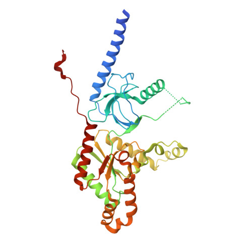
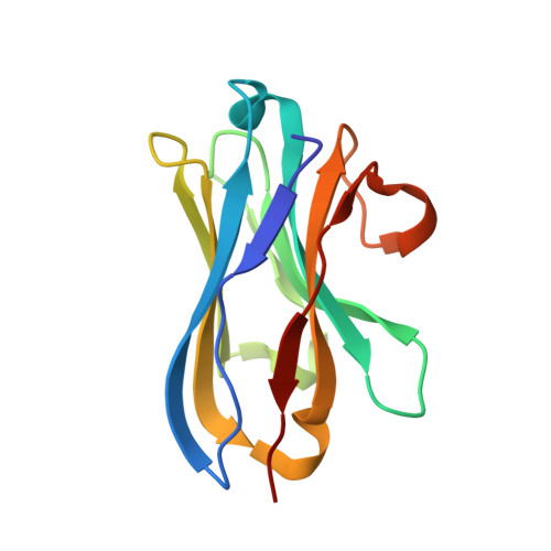
Selective posttranslational inhibition of Ca V beta 1 -associated voltage-dependent calcium channels with a functionalized nanobody.

Morgenstern, T.J.Nirwan, N.Hernandez-Ochoa, E.O.Bibollet, H.Choudhury, P.Laloudakis, Y.D.Ben Johny, M.Bannister, R.A.Schneider, M.F.Minor Jr., D.L.Colecraft, H.M.

(2022) Nat Commun 13: 7556-7556

  • DOI: https://doi.org/10.1038/s41467-022-35025-7
  • Primary Citation Related Structures: 
    8DAM, 8E0E

  • PubMed Abstract: 

    Ca 2+ influx through high-voltage-activated calcium channels (HVACCs) controls diverse cellular functions. A critical feature enabling a singular signal, Ca 2+ influx, to mediate disparate functions is diversity of HVACC pore-forming α 1 and auxiliary Ca V β 1 -Ca V β 4 subunits. Selective Ca V α 1 blockers have enabled deciphering their unique physiological roles. By contrast, the capacity to post-translationally inhibit HVACCs based on Ca V β isoform is non-existent. Conventional gene knockout/shRNA approaches do not adequately address this deficit owing to subunit reshuffling and partially overlapping functions of Ca V β isoforms. Here, we identify a nanobody (nb.E8) that selectively binds Ca V β 1 SH3 domain and inhibits Ca V β 1 -associated HVACCs by reducing channel surface density, decreasing open probability, and speeding inactivation. Functionalizing nb.E8 with Nedd4L HECT domain yielded Chisel-1 which eliminated current through Ca V β 1 -reconstituted Ca V 1/Ca V 2 and native Ca V 1.1 channels in skeletal muscle, strongly suppressed depolarization-evoked Ca 2+ influx and excitation-transcription coupling in hippocampal neurons, but was inert against Ca V β 2 -associated Ca V 1.2 in cardiomyocytes. The results introduce an original method for probing distinctive functions of ion channel auxiliary subunit isoforms, reveal additional dimensions of Ca V β 1 signaling in neurons, and describe a genetically-encoded HVACC inhibitor with unique properties.


  • Organizational Affiliation
    • Department of Molecular Pharmacology and Therapeutics, Columbia University Irving Medical Center, New York, NY, USA.

Macromolecule Content 

  • Total Structure Weight: 53.72 kDa 
  • Atom Count: 3,787 
  • Modeled Residue Count: 443 
  • Deposited Residue Count: 471 
  • Unique protein chains: 2

Macromolecules

Find similar proteins by:|  3D Structure
Entity ID: 1
MoleculeChains  Sequence LengthOrganismDetailsImage
Voltage-dependent L-type calcium channel subunit beta-2349Rattus norvegicusMutation(s): 0 
Gene Names: Cacnb2
Find similar proteins by:|  3D Structure
Entity ID: 2
MoleculeChains  Sequence LengthOrganismDetailsImage
Nanobody nb.F3122Lama glamaMutation(s): 0 

Small Molecules

Ligands 4 Unique
IDChains Name / Formula / InChI Key2D Diagram3D Interactions
MPD

Query on MPD


Download:Ideal Coordinates CCD File
D [auth A],
G [auth A],
H [auth A],
J [auth A]
(4S)-2-METHYL-2,4-PENTANEDIOL
C6 H14 O2
SVTBMSDMJJWYQN-YFKPBYRVSA-N
GOL

Query on GOL


Download:Ideal Coordinates CCD File
C [auth A],
E [auth A],
F [auth A],
I [auth A],
K [auth A]
GLYCEROL
C3 H8 O3
PEDCQBHIVMGVHV-UHFFFAOYSA-N
CA

Query on CA


Download:Ideal Coordinates CCD File
L [auth A]CALCIUM ION
Ca
BHPQYMZQTOCNFJ-UHFFFAOYSA-N
MG

Query on MG


Download:Ideal Coordinates CCD File
M [auth A]MAGNESIUM ION
Mg
JLVVSXFLKOJNIY-UHFFFAOYSA-N

Experimental Data & Validation

Experimental Data

  • Method: X-RAY DIFFRACTION
  • Resolution: 2.00 Å
  • R-Value Free:  0.221 (Depositor), 0.197 (DCC) 
  • R-Value Work:  0.197 (Depositor), 0.197 (DCC) 
  • R-Value Observed: 0.199 (Depositor) 
Space Group: P 65
Unit Cell:
Length ( Å )Angle ( ˚ )
a = 144.525α = 90
b = 144.525β = 90
c = 63.596γ = 120
Software Package:
Software NamePurpose
Aimlessdata scaling
PHENIXrefinement
PDB_EXTRACTdata extraction
XDSdata reduction
PHASERphasing

Structure Validation

View Full Validation Report


Entry History 

& Funding Information

Deposition Data


Funding OrganizationLocationGrant Number
National Institutes of Health/National Heart, Lung, and Blood Institute (NIH/NHLBI)United States--

Revision History  (Full details and data files)

  • Version 1.0: 2023-01-18
    Type: Initial release
  • Version 1.1: 2023-10-25
    Changes: Data collection, Refinement description