8DU3 | pdb_00008du3

Crystal structure of A2AAR-StaR2-bRIL in complex with compound 21a


Experimental Data Snapshot

  • Method: X-RAY DIFFRACTION
  • Resolution: 2.50 Å
  • R-Value Free: 
    0.232 (Depositor), 0.235 (DCC) 
  • R-Value Work: 
    0.189 (Depositor), 0.196 (DCC) 
  • R-Value Observed: 
    0.191 (Depositor) 

Starting Model: experimental
View more details

wwPDB Validation 3D Report Full Report

Validation slider image for 8DU3

Ligand Structure Quality Assessment 


This is version 1.3 of the entry. See complete history

Literature

High ligand efficiency quinazoline compounds as novel A 2A adenosine receptor antagonists.

Bolteau, R.Duroux, R.Laversin, A.Vreulz, B.Shiriaeva, A.Stauch, B.Han, G.W.Cherezov, V.Renault, N.Barczyk, A.Ravez, S.Coevoet, M.Melnyk, P.Liberelle, M.Yous, S.

(2022) Eur J Med Chem 241: 114620-114620

  • DOI: https://doi.org/10.1016/j.ejmech.2022.114620
  • Primary Citation Related Structures: 
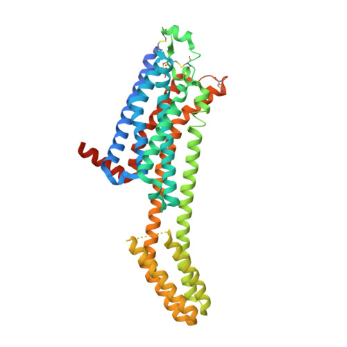
    8DU3

  • PubMed Abstract: 

    The past fifty years have been marked by the surge of neurodegenerative diseases. Unfortunately, current treatments are only symptomatic. Hence, the search for new and innovative therapeutic targets for curative treatments becomes a major challenge. Among these targets, the adenosine A 2A receptor (A 2A AR) has been the subject of much research in recent years. In this paper, we report the design, synthesis and pharmacological analysis of quinazoline derivatives as A 2A AR antagonists with high ligand efficiency. This class of molecules has been discovered by a virtual screening and bears no structural semblance with reference antagonist ZM-241385. More precisely, we identified a series of 2-aminoquinazoline as promising A 2A AR antagonists. Among them, one compound showed a high affinity towards A 2A AR (21a, K i  = 20 nM). We crystallized this ligand in complex with A 2A AR, confirming one of our predicted docking poses and opening up possibilities for further optimization to derive selective ligands for specific adenosine receptor subtypes.


  • Organizational Affiliation
    • Univ. Lille, Inserm, CHU Lille, U1172, LilNCog - Lille Neuroscience & Cognition, F-59000, Lille, France.

Macromolecule Content 

  • Total Structure Weight: 57.35 kDa 
  • Atom Count: 3,466 
  • Modeled Residue Count: 390 
  • Deposited Residue Count: 447 
  • Unique protein chains: 1

Macromolecules

Find similar proteins by:|  3D Structure
Entity ID: 1
MoleculeChains  Sequence LengthOrganismDetailsImage
Adenosine receptor A2a, soluble cytochrome b562 chimera,Soluble cytochrome b562,Adenosine receptor A2a447Homo sapiensMutation(s): 12 
Gene Names: ADORA2AADORA2cybC
Membrane Entity: Yes 
UniProt & NIH Common Fund Data Resources
Find proteins for P0ABE7 (Escherichia coli)
Explore P0ABE7 
Go to UniProtKB:  P0ABE7
Find proteins for P29274 (Homo sapiens)
Explore P29274 
Go to UniProtKB:  P29274
PHAROS:  P29274
GTEx:  ENSG00000128271 
Entity Groups
Sequence Clusters30% Identity50% Identity70% Identity90% Identity95% Identity100% Identity
UniProt GroupsP0ABE7P29274
Sequence Annotations
Expand
Reference Sequence

Small Molecules

Ligands 4 Unique
IDChains Name / Formula / InChI Key2D Diagram3D Interactions
CLR

Query on CLR


Download:Ideal Coordinates CCD File
C [auth A],
D [auth A],
E [auth A]
CHOLESTEROL
C27 H46 O
HVYWMOMLDIMFJA-DPAQBDIFSA-N
OLC

Query on OLC


Download:Ideal Coordinates CCD File
F [auth A],
G [auth A],
H [auth A],
I [auth A]
(2R)-2,3-dihydroxypropyl (9Z)-octadec-9-enoate
C21 H40 O4
RZRNAYUHWVFMIP-GDCKJWNLSA-N
TKO
(Subject of Investigation/LOI)

Query on TKO


Download:Ideal Coordinates CCD File
B [auth A](4M)-6-bromo-4-(furan-2-yl)quinazolin-2-amine
C12 H8 Br N3 O
XDIROSBRBKJBMH-UHFFFAOYSA-N
OLA

Query on OLA


Download:Ideal Coordinates CCD File
J [auth A]
K [auth A]
L [auth A]
M [auth A]
N [auth A]
J [auth A],
K [auth A],
L [auth A],
M [auth A],
N [auth A],
O [auth A],
P [auth A],
Q [auth A],
R [auth A],
S [auth A],
T [auth A],
U [auth A],
V [auth A],
W [auth A],
X [auth A],
Y [auth A],
Z [auth A]
OLEIC ACID
C18 H34 O2
ZQPPMHVWECSIRJ-KTKRTIGZSA-N

Experimental Data & Validation

Experimental Data

  • Method: X-RAY DIFFRACTION
  • Resolution: 2.50 Å
  • R-Value Free:  0.232 (Depositor), 0.235 (DCC) 
  • R-Value Work:  0.189 (Depositor), 0.196 (DCC) 
  • R-Value Observed: 0.191 (Depositor) 
Space Group: C 2 2 21
Unit Cell:
Length ( Å )Angle ( ˚ )
a = 39.6α = 90
b = 179.8β = 90
c = 141γ = 90
Software Package:
Software NamePurpose
PHENIXrefinement
PDB_EXTRACTdata extraction
HKL-2000data reduction
HKL-2000data scaling
PHASERphasing

Structure Validation

View Full Validation Report


Ligand Structure Quality Assessment 


Entry History 

& Funding Information

Deposition Data


Funding OrganizationLocationGrant Number
National Institutes of Health/National Cancer Institute (NIH/NCI)United StatesACB-12002

Revision History  (Full details and data files)

  • Version 1.0: 2022-08-10
    Type: Initial release
  • Version 1.1: 2022-08-24
    Changes: Database references
  • Version 1.2: 2023-10-18
    Changes: Data collection, Refinement description
  • Version 1.3: 2024-10-16
    Changes: Structure summary