8DSJ

Peptidylglycine alpha hydroxylating monooxygenase anaerobic


Experimental Data Snapshot

  • Method: X-RAY DIFFRACTION
  • Resolution: 2.80 Å
  • R-Value Free: 0.275 
  • R-Value Work: 0.197 
  • R-Value Observed: 0.201 

wwPDB Validation   3D Report Full Report


This is version 1.3 of the entry. See complete history


Literature

New structures reveal flexible dynamics between the subdomains of peptidylglycine monooxygenase. Implications for an open to closed mechanism.

Arias, R.J.Welch, E.F.Blackburn, N.J.

(2023) Protein Sci 32: e4615-e4615

  • DOI: https://doi.org/10.1002/pro.4615
  • Primary Citation of Related Structures:  
    8DSJ, 8DSL, 8DSN

  • PubMed Abstract: 

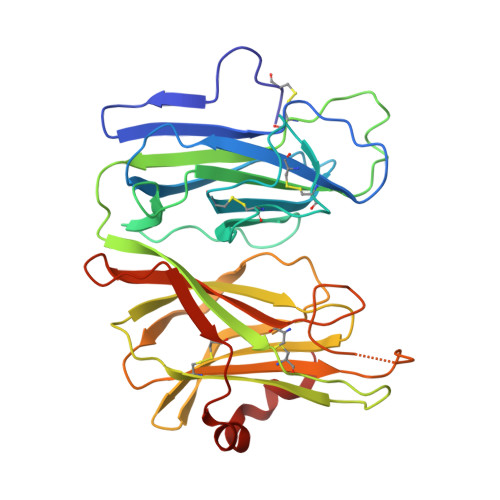
    Peptidylglycine monooxygenase (PHM) is essential for the biosynthesis of many neuroendocrine peptides via a copper-dependent hydroxylation of a glycine-extended pro-peptide. The "canonical" mechanism requires the transfer of two electrons from one mononuclear copper (CuH, H-site) to a second mononuclear copper (CuM, M-site) which is the site of oxygen binding and catalysis. In most crystal structures the copper centers are separated by 11 Å of disordered solvent, but recent work has established that a PHM variant H108A forms a closed conformer in the presence of citrate with a reduced Cu-Cu site separation of ~4 Å. Here we report three new PHM structures where the H and M sites are separated by a longer distance of ~14 Å. Variation in Cu-Cu distance is the result of a rotation of the M subdomain about a hinge point centered on the pro 199 -leu 200 -ile 201 triad which forms the linker between subdomains. The energetic cost of domain dynamics is likely small enough to allow free rotation of the subdomains relative to each other, adding credence to recent suggestions that an open-to-closed transition to form a binuclear oxygen binding intermediate is an essential element of catalysis. This inference would explain many experimental observations that are inconsistent with the current canonical mechanism including substrate-induced oxygen activation and isotope scrambling during the peroxide shunt.


  • Organizational Affiliation

    Department of Chemical Physiology and Biochemistry, Oregon Health and Science University, Portland, Oregon, USA.


Macromolecules
Find similar proteins by:  (by identity cutoff)  |  3D Structure
Entity ID: 1
MoleculeChains Sequence LengthOrganismDetailsImage
Peptidylglycine alpha-amidating monooxygenaseA,
B [auth K]
310Rattus norvegicusMutation(s): 0 
Gene Names: Pam
EC: 1.14.17.3 (PDB Primary Data), 4.3.2.5 (PDB Primary Data)
UniProt
Find proteins for P14925 (Rattus norvegicus)
Explore P14925 
Go to UniProtKB:  P14925
Entity Groups  
Sequence Clusters30% Identity50% Identity70% Identity90% Identity95% Identity100% Identity
UniProt GroupP14925
Sequence Annotations
Expand
  • Reference Sequence
Small Molecules
Ligands 2 Unique
IDChains Name / Formula / InChI Key2D Diagram3D Interactions
GOL
Query on GOL

Download Ideal Coordinates CCD File 
F [auth A],
G [auth A],
K,
L [auth K],
M [auth K]
GLYCEROL
C3 H8 O3
PEDCQBHIVMGVHV-UHFFFAOYSA-N
CU (Subject of Investigation/LOI)
Query on CU

Download Ideal Coordinates CCD File 
C [auth A]
D [auth A]
E [auth A]
H [auth K]
I [auth K]
C [auth A],
D [auth A],
E [auth A],
H [auth K],
I [auth K],
J [auth K]
COPPER (II) ION
Cu
JPVYNHNXODAKFH-UHFFFAOYSA-N
Experimental Data & Validation

Experimental Data

  • Method: X-RAY DIFFRACTION
  • Resolution: 2.80 Å
  • R-Value Free: 0.275 
  • R-Value Work: 0.197 
  • R-Value Observed: 0.201 
  • Space Group: P 1
Unit Cell:
Length ( Å )Angle ( ˚ )
a = 38.14α = 84.85
b = 53.263β = 89.96
c = 86.424γ = 78.5
Software Package:
Software NamePurpose
Aimlessdata scaling
REFMACrefinement
PDB_EXTRACTdata extraction
XDSdata reduction
MOLREPphasing

Structure Validation

View Full Validation Report


Entry History & Funding Information

Deposition Data


Funding OrganizationLocationGrant Number
National Institutes of Health/National Institute of General Medical Sciences (NIH/NIGMS)United StatesR35 GM136239

Revision History  (Full details and data files)

  • Version 1.0: 2023-03-22
    Type: Initial release
  • Version 1.1: 2023-04-12
    Changes: Database references
  • Version 1.2: 2023-04-19
    Changes: Database references
  • Version 1.3: 2023-10-25
    Changes: Data collection, Refinement description