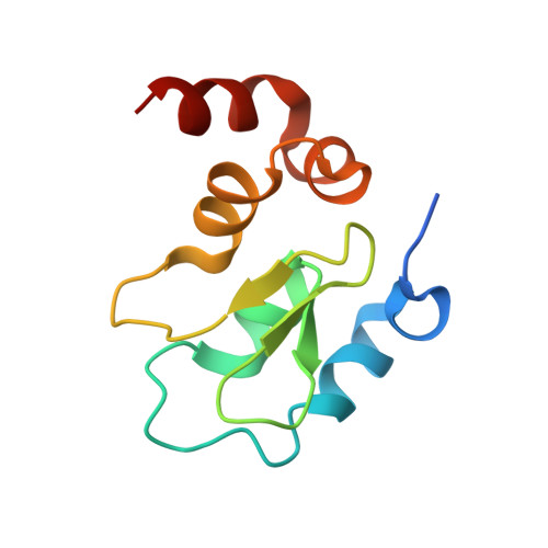
8DSF

Structure of cIAP1 with BCCov


Experimental Data Snapshot

  • Method: X-RAY DIFFRACTION
  • Resolution: 1.50 Å
  • R-Value Free: 0.227 
  • R-Value Work: 0.189 
  • R-Value Observed: 0.191 

wwPDB Validation   3D Report Full Report


Ligand Structure Quality Assessment 


This is version 1.2 of the entry. See complete history


Literature

A covalent BTK ternary complex compatible with targeted protein degradation.

Schiemer, J.Maxwell, A.Horst, R.Liu, S.Uccello, D.P.Borzilleri, K.Rajamohan, N.Brown, M.F.Calabrese, M.F.

(2023) Nat Commun 14: 1189-1189

  • DOI: https://doi.org/10.1038/s41467-023-36738-z
  • Primary Citation of Related Structures:  
    8DSF, 8DSO

  • PubMed Abstract: 

    Targeted protein degradation using heterobifunctional chimeras holds the potential to expand target space and grow the druggable proteome. Most acutely, this provides an opportunity to target proteins that lack enzymatic activity or have otherwise proven intractable to small molecule inhibition. Limiting this potential, however, is the remaining need to develop a ligand for the target of interest. While a number of challenging proteins have been successfully targeted by covalent ligands, unless this modification affects form or function, it may lack the ability to drive a biological response. Bridging covalent ligand discovery with chimeric degrader design has emerged as a potential mechanism to advance both fields. In this work, we employ a set of biochemical and cellular tools to deconvolute the role of covalent modification in targeted protein degradation using Bruton's tyrosine kinase. Our results reveal that covalent target modification is fundamentally compatible with the protein degrader mechanism of action.


  • Organizational Affiliation

    Discovery Sciences, Pfizer Worldwide Research and Development, Groton, CT, USA.


Macromolecules
Find similar proteins by:  (by identity cutoff)  |  3D Structure
Entity ID: 1
MoleculeChains Sequence LengthOrganismDetailsImage
Baculoviral IAP repeat-containing protein 2
A, B, C, D
99Homo sapiensMutation(s): 0 
Gene Names: BIRC2API1MIHBRNF48
EC: 2.3.2.27
UniProt & NIH Common Fund Data Resources
Find proteins for Q13490 (Homo sapiens)
Explore Q13490 
Go to UniProtKB:  Q13490
PHAROS:  Q13490
GTEx:  ENSG00000110330 
Entity Groups  
Sequence Clusters30% Identity50% Identity70% Identity90% Identity95% Identity100% Identity
UniProt GroupQ13490
Sequence Annotations
Expand
  • Reference Sequence
Small Molecules
Ligands 2 Unique
IDChains Name / Formula / InChI Key2D Diagram3D Interactions
TO0 (Subject of Investigation/LOI)
Query on TO0

Download Ideal Coordinates CCD File 
F [auth A],
H [auth B],
J [auth C],
L [auth D]
(4S)-4-[2-(2-{4-[(2E)-4-{(3R)-3-[4-amino-3-(4-phenoxyphenyl)-1H-pyrazolo[3,4-d]pyrimidin-1-yl]piperidin-1-yl}-4-oxobut-2-en-1-yl]piperazin-1-yl}ethoxy)acetamido]-1-{(2S)-2-cyclohexyl-2-[(N-methyl-L-alanyl)amino]acetyl}-N-[(1R)-1,2,3,4-tetrahydronaphthalen-1-yl]-L-prolinamide unbound form
C61 H79 N13 O7
TVZVBEZZTQUFMH-HRIQDGSKSA-N
ZN
Query on ZN

Download Ideal Coordinates CCD File 
E [auth A],
G [auth B],
I [auth C],
K [auth D]
ZINC ION
Zn
PTFCDOFLOPIGGS-UHFFFAOYSA-N
Experimental Data & Validation

Experimental Data

  • Method: X-RAY DIFFRACTION
  • Resolution: 1.50 Å
  • R-Value Free: 0.227 
  • R-Value Work: 0.189 
  • R-Value Observed: 0.191 
  • Space Group: P 1
Unit Cell:
Length ( Å )Angle ( ˚ )
a = 28.99α = 96.7
b = 30.77β = 90.22
c = 118.63γ = 111.08
Software Package:
Software NamePurpose
BUSTERrefinement
PDB_EXTRACTdata extraction
autoPROCdata reduction
autoPROCdata scaling
PHASERphasing

Structure Validation

View Full Validation Report


Ligand Structure Quality Assessment 


Entry History & Funding Information

Deposition Data


Funding OrganizationLocationGrant Number
Not funded--

Revision History  (Full details and data files)

  • Version 1.0: 2023-03-08
    Type: Initial release
  • Version 1.1: 2023-10-25
    Changes: Data collection, Refinement description
  • Version 1.2: 2024-05-01
    Changes: Database references