


Local refinement of SARS-CoV-2 vaccine induced antibody Ab026500 bound to SARS-CoV-2 HexaPro RBD Spike ectodomain
Stalls, V., Acharya, P., May, A.J., Malewana, D.To be published.
Experimental Data Snapshot
Starting Models: experimental
View more details
wwPDB Validation 3D Report Full Report

Entity ID: 1 | |||||
|---|---|---|---|---|---|
| Molecule | Chains | Sequence Length | Organism | Details | Image |
| Spike protein S1 | A [auth B] | 205 | Severe acute respiratory syndrome coronavirus 2 | Mutation(s): 0 Gene Names: S, 2 | 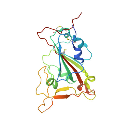 |
UniProt | |||||
Entity Groups | |||||
| Sequence Clusters | 30% Identity50% Identity70% Identity90% Identity95% Identity100% Identity | ||||
| UniProt Group | P0DTC2 | ||||
Glycosylation | |||||
| Glycosylation Sites: 1 | Go to GlyGen: P0DTC2-1 | ||||
Sequence AnnotationsExpand | |||||
Reference Sequence | |||||
Entity ID: 2 | |||||
|---|---|---|---|---|---|
| Molecule | Chains | Sequence Length | Organism | Details | Image |
| DH1338 Fab Heavy Chain | B [auth F] | 128 | Macaca mulatta | Mutation(s): 0 | 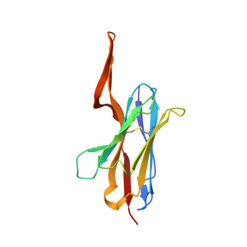 |
Entity ID: 3 | |||||
|---|---|---|---|---|---|
| Molecule | Chains | Sequence Length | Organism | Details | Image |
| DH1338 Fab Light Chain | C [auth G] | 107 | Macaca mulatta | Mutation(s): 0 | 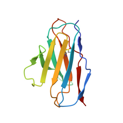 |
| Task | Software Package | Version |
|---|---|---|
| RECONSTRUCTION | Coot | |
| RECONSTRUCTION | ISOLDE | |
| RECONSTRUCTION | PHENIX | |
| MODEL REFINEMENT | PHENIX |
| Funding Organization | Location | Grant Number |
|---|---|---|
| National Institutes of Health/National Institute Of Allergy and Infectious Diseases (NIH/NIAID) | United States | R01 AI165947 |


