8DLA

ClpP2 from Chlamydia trachomatis bound by MAS1-12


Experimental Data Snapshot

  • Method: X-RAY DIFFRACTION
  • Resolution: 2.66 Å
  • R-Value Free: 0.210 
  • R-Value Work: 0.173 
  • R-Value Observed: 0.175 

wwPDB Validation   3D Report Full Report


Ligand Structure Quality Assessment 


This is version 1.1 of the entry. See complete history


Literature

The structure of caseinolytic protease subunit ClpP2 reveals a functional model of the caseinolytic protease system from Chlamydia trachomatis.

Azadmanesh, J.Seleem, M.A.Struble, L.Wood, N.A.Fisher, D.J.Lovelace, J.J.Artigues, A.Fenton, A.W.Borgstahl, G.E.O.Ouellette, S.P.Conda-Sheridan, M.

(2023) J Biol Chem 299: 102762-102762

  • DOI: https://doi.org/10.1016/j.jbc.2022.102762
  • Primary Citation of Related Structures:  
    8DLA

  • PubMed Abstract: 

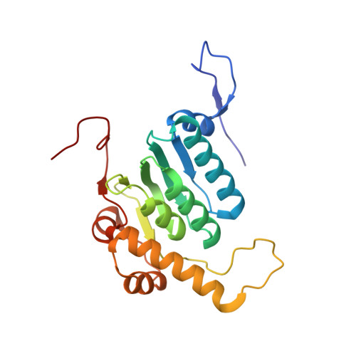
    Chlamydia trachomatis (ct) is the most reported bacterial sexually transmitted infection worldwide and the leading cause of preventable blindness. Caseinolytic proteases (ClpP) from pathogenic bacteria are attractive antibiotic targets, particularly for bacterial species that form persister colonies with phenotypic resistance against common antibiotics. ClpP functions as a multisubunit proteolytic complex, and bacteria are eradicated when ClpP is disrupted. Although crucial for chlamydial development and the design of agents to treat chlamydia, the structures of ctClpP1 and ctClpP2 have yet to be solved. Here, we report the first crystal structure of full-length ClpP2 as an inactive homotetradecamer in a complex with a candidate antibiotic at 2.66 Å resolution. The structure details the functional domains of the ClpP2 protein subunit and includes the handle domain, which is integral to proteolytic activation. In addition, hydrogen-deuterium exchange mass spectroscopy probed the dynamics of ClpP2, and molecular modeling of ClpP1 predicted an assembly with ClpP2. By leveraging previous enzymatic experiments, we constructed a model of ClpP2 activation and its interaction with the protease subunits ClpP1 and ClpX. The structural information presented will be relevant for future rational drug design against these targets and will lead to a better understanding of ClpP complex formation and activation within this important human pathogen.


  • Organizational Affiliation

    The Eppley Institute for Research in Cancer and Allied Diseases, Fred & Pamela Buffett Cancer Center, University of Nebraska Medical Center, Omaha, Nebraska, USA.


Macromolecules
Find similar proteins by:  (by identity cutoff)  |  3D Structure
Entity ID: 1
MoleculeChains Sequence LengthOrganismDetailsImage
ATP-dependent Clp protease proteolytic subunit 2
A, B, C, D, E
A, B, C, D, E, F, G, H, I, J, K, L, M, N
203Chlamydia trachomatisMutation(s): 0 
Gene Names: clpP2
EC: 3.4.21.92
UniProt
Find proteins for O84712 (Chlamydia trachomatis (strain D/UW-3/Cx))
Explore O84712 
Go to UniProtKB:  O84712
Entity Groups  
Sequence Clusters30% Identity50% Identity70% Identity90% Identity95% Identity100% Identity
UniProt GroupO84712
Sequence Annotations
Expand
  • Reference Sequence
Small Molecules
Ligands 3 Unique
IDChains Name / Formula / InChI Key2D Diagram3D Interactions
UER (Subject of Investigation/LOI)
Query on UER

Download Ideal Coordinates CCD File 
DA [auth M]
O [auth A]
P [auth A]
Q [auth B]
U [auth D]
DA [auth M],
O [auth A],
P [auth A],
Q [auth B],
U [auth D],
Z [auth G]
1-{4-[(4-chlorophenyl)methyl]piperazin-1-yl}-2-methyl-2-[5-(trifluoromethyl)pyridine-2-sulfonyl]propan-1-one
C21 H23 Cl F3 N3 O3 S
MFQQEGNFXSEGTC-UHFFFAOYSA-N
GOL
Query on GOL

Download Ideal Coordinates CCD File 
EA [auth N],
S [auth B],
T [auth C],
Y [auth G]
GLYCEROL
C3 H8 O3
PEDCQBHIVMGVHV-UHFFFAOYSA-N
NA
Query on NA

Download Ideal Coordinates CCD File 
AA [auth G]
BA [auth H]
CA [auth L]
FA [auth N]
R [auth B]
AA [auth G],
BA [auth H],
CA [auth L],
FA [auth N],
R [auth B],
V [auth D],
W [auth E],
X [auth F]
SODIUM ION
Na
FKNQFGJONOIPTF-UHFFFAOYSA-N
Experimental Data & Validation

Experimental Data

  • Method: X-RAY DIFFRACTION
  • Resolution: 2.66 Å
  • R-Value Free: 0.210 
  • R-Value Work: 0.173 
  • R-Value Observed: 0.175 
  • Space Group: P 1
Unit Cell:
Length ( Å )Angle ( ˚ )
a = 97α = 97.16
b = 98.01β = 114.15
c = 97.97γ = 114.16
Software Package:
Software NamePurpose
PHENIXrefinement
MOSFLMdata reduction
Aimlessdata scaling
PHENIXphasing

Structure Validation

View Full Validation Report


Ligand Structure Quality Assessment 


Entry History & Funding Information

Deposition Data


Funding OrganizationLocationGrant Number
National Institutes of Health/National Institute of General Medical Sciences (NIH/NIGMS)United States1P20GM121316-01A1
Department of Defense (DOD, United States)United StatesPRMRP#PR172445
National Institutes of Health/National Cancer Institute (NIH/NCI)United States5P30CA036727-33

Revision History  (Full details and data files)

  • Version 1.0: 2023-08-09
    Type: Initial release
  • Version 1.1: 2024-01-24
    Changes: Data collection, Database references