8DJK

HMGCR-UBIAD1 Complex State 2


Experimental Data Snapshot

  • Method: ELECTRON MICROSCOPY
  • Resolution: 3.33 Å
  • Aggregation State: PARTICLE 
  • Reconstruction Method: SINGLE PARTICLE 

wwPDB Validation   3D Report Full Report


This is version 1.0 of the entry. See complete history


Literature

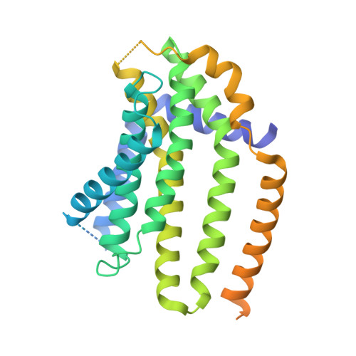
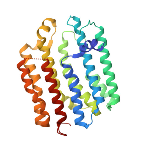
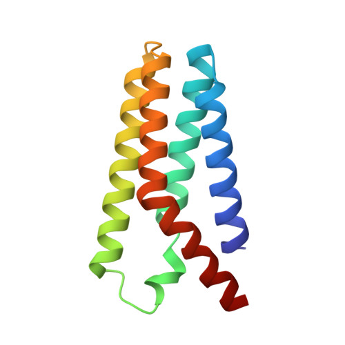
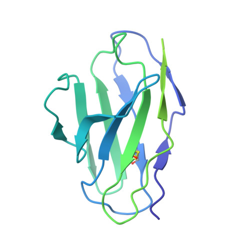
Regulated degradation of HMG CoA reductase requires conformational changes in sterol-sensing domain.

Chen, H.Qi, X.Faulkner, R.A.Schumacher, M.M.Donnelly, L.M.DeBose-Boyd, R.A.Li, X.

(2022) Nat Commun 13: 4273-4273

  • DOI: https://doi.org/10.1038/s41467-022-32025-5
  • Primary Citation of Related Structures:  
    8DJK, 8DJM

  • PubMed Abstract: 

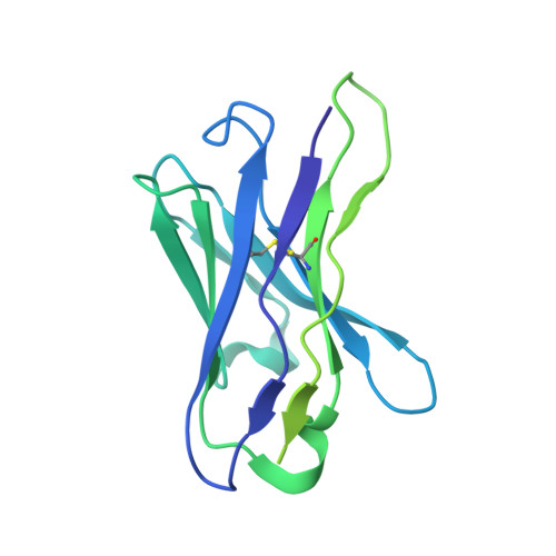
    3-Hydroxy-3-methylglutaryl coenzyme A reductase (HMGCR) is the rate-limiting enzyme in cholesterol synthesis and target of cholesterol-lowering statin drugs. Accumulation of sterols in endoplasmic reticulum (ER) membranes accelerates degradation of HMGCR, slowing the synthesis of cholesterol. Degradation of HMGCR is inhibited by its binding to UBIAD1 (UbiA prenyltransferase domain-containing protein-1). This inhibition contributes to statin-induced accumulation of HMGCR, which limits their cholesterol-lowering effects. Here, we report cryo-electron microscopy structures of the HMGCR-UBIAD1 complex, which is maintained by interactions between transmembrane helix (TM) 7 of HMGCR and TMs 2-4 of UBIAD1. Disrupting this interface by mutagenesis prevents complex formation, enhancing HMGCR degradation. TMs 2-6 of HMGCR contain a 170-amino acid sterol sensing domain (SSD), which exists in two conformations-one of which is essential for degradation. Thus, our data supports a model that rearrangement of the TMs in the SSD permits recruitment of proteins that initate HMGCR degradation, a key reaction in the regulatory system that governs cholesterol synthesis.


  • Organizational Affiliation

    Department of Molecular Genetics, University of Texas Southwestern Medical Center, Dallas, TX, USA.


Macromolecules
Find similar proteins by:  (by identity cutoff)  |  3D Structure
Entity ID: 1
MoleculeChains Sequence LengthOrganismDetailsImage
3-hydroxy-3-methylglutaryl-coenzyme A reductase364Cricetulus griseusMutation(s): 0 
Gene Names: HMGCR
EC: 1.1.1.34
Membrane Entity: Yes 
UniProt
Find proteins for P00347 (Cricetulus griseus)
Explore P00347 
Go to UniProtKB:  P00347
Entity Groups  
Sequence Clusters30% Identity50% Identity70% Identity90% Identity95% Identity100% Identity
UniProt GroupP00347
Sequence Annotations
Expand
  • Reference Sequence
Find similar proteins by:  (by identity cutoff)  |  3D Structure
Entity ID: 2
MoleculeChains Sequence LengthOrganismDetailsImage
UbiA prenyltransferase domain-containing protein 1301Cricetulus griseusMutation(s): 0 
Gene Names: Ubiad1CgPICR_017873H671_2g6271I79_022097
Membrane Entity: Yes 
UniProt
Find proteins for G3IEF0 (Cricetulus griseus)
Explore G3IEF0 
Go to UniProtKB:  G3IEF0
Entity Groups  
Sequence Clusters30% Identity50% Identity70% Identity90% Identity95% Identity100% Identity
UniProt GroupG3IEF0
Sequence Annotations
Expand
  • Reference Sequence
Find similar proteins by:  (by identity cutoff)  |  3D Structure
Entity ID: 3
MoleculeChains Sequence LengthOrganismDetailsImage
Soluble cytochrome b562120Escherichia coliMutation(s): 0 
Gene Names: cybC
Membrane Entity: Yes 
UniProt
Find proteins for P0ABE7 (Escherichia coli)
Explore P0ABE7 
Go to UniProtKB:  P0ABE7
Entity Groups  
Sequence Clusters30% Identity50% Identity70% Identity90% Identity95% Identity100% Identity
UniProt GroupP0ABE7
Sequence Annotations
Expand
  • Reference Sequence
Find similar proteins by:  (by identity cutoff)  |  3D Structure
Entity ID: 4
MoleculeChains Sequence LengthOrganismDetailsImage
Fab 15B2 Light ChainD [auth L]214Mus musculusMutation(s): 0 
Entity Groups  
Sequence Clusters30% Identity50% Identity70% Identity90% Identity95% Identity100% Identity
Sequence Annotations
Expand
  • Reference Sequence
Find similar proteins by:  (by identity cutoff)  |  3D Structure
Entity ID: 5
MoleculeChains Sequence LengthOrganismDetailsImage
Fab 15B2 Heavy ChainE [auth H]232Mus musculusMutation(s): 0 
Entity Groups  
Sequence Clusters30% Identity50% Identity70% Identity90% Identity95% Identity100% Identity
Sequence Annotations
Expand
  • Reference Sequence
Experimental Data & Validation

Experimental Data

  • Method: ELECTRON MICROSCOPY
  • Resolution: 3.33 Å
  • Aggregation State: PARTICLE 
  • Reconstruction Method: SINGLE PARTICLE 

Structure Validation

View Full Validation Report


Entry History & Funding Information

Deposition Data


Funding OrganizationLocationGrant Number
National Institutes of Health/National Institute of General Medical Sciences (NIH/NIGMS)United States--
Welch FoundationUnited States--

Revision History  (Full details and data files)

  • Version 1.0: 2022-08-03
    Type: Initial release