The Y430F mutant of Salmonella d-ornithine/d-lysine decarboxylase has altered stereospecificity and a putrescine allosteric activation site.
Phillips, R.S., Nguyen Hoang, K.N.(2022) Arch Biochem Biophys 731: 109429-109429
- PubMed: 36265649 Search on PubMedSearch on PubMed Central
- DOI: https://doi.org/10.1016/j.abb.2022.109429
- Primary Citation Related Structures:
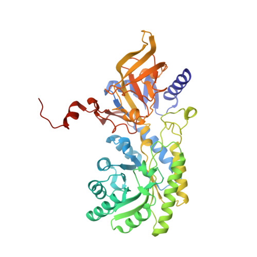
8D2Y, 8D4I, 8D5D, 8D5R, 8D88 - PubMed Abstract:
Tyrosine-430 of d-ornithine/d-lysine decarboxylase (DOKDC) is located in the active site, and was suggested to be responsible for the D-stereospecificity of the enzyme. We have prepared the Y430F mutant form of Salmonella enterica serovar typhimurium DOKDC and evaluated its catalytic activity with D- and l-lysine and ornithine. The kinetic results show that the Y430F mutant has measurable decarboxylase activity with both D- and l-lysine and ornithine, which wild type DOKDC does not. Spectroscopic experiments show that these amino acids bind to form external aldimine complexes with the pyridoxal-5'-phosphate with λ max = 425 nm. In addition, we have obtained crystal structures of Y430F DOKDC bound to HEPES, putrescine, d-ornithine, d-lysine, and d-arginine. The d-amino acids bind in the crystals to form equilibrium mixtures of gem-diamine and external aldimine complexes. Furthermore, the crystal structures reveal an unexpected allosteric product activator site for putrescine located on the 2-fold axis between the two active sites. Putrescine binds by donating hydrogen bonds from the ammonium groups to Asp-361 and Gln-358 in the specificity helix of both chains. Addition of 0.1-1 mM putrescine eliminates the lag in steady state kinetics and abolishes the sigmoid kinetics. The catalytic loop was modeled with AlphaFold2, and the model shows that Glu-181 can form additional hydrogen bonds with the bound putrescine, likely stabilizing the catalytic closed conformation.
- Department of Chemistry, University of Georgia, Athens, GA, 30602, USA; Department of Biochemistry and Molecular Biology, University of Georgia, Athens, GA, 30602, USA. Electronic address: plp@uga.edu.
Organizational Affiliation: