Selective Oxidations Using a Cytochrome P450 Enzyme Variant Driven with Surrogate Oxygen Donors and Light.
Lee, J.H.Z., Podgorski, M.N., Moir, M., Gee, A.R., Bell, S.G.(2022) Chemistry 28: e202201366-e202201366
- PubMed: 35712785 Search on PubMedSearch on PubMed Central
- DOI: https://doi.org/10.1002/chem.202201366
- Primary Citation Related Structures:
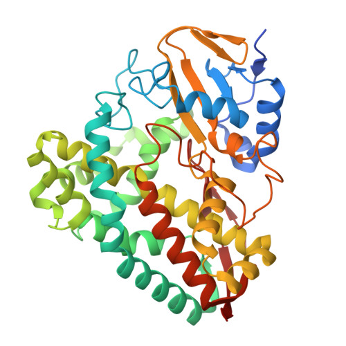
8D1C - PubMed Abstract:
Cytochrome P450 monooxygenase enzymes are versatile catalysts, which have been adapted for multiple applications in chemical synthesis. Mutation of a highly conserved active site threonine to a glutamate can convert these enzymes into peroxygenases that utilise hydrogen peroxide (H 2 O 2 ). Here, we use the T252E-CYP199A4 variant to study peroxide-driven oxidation activity by using H 2 O 2 and urea-hydrogen peroxide (UHP). We demonstrate that the T252E variant has a higher stability to H 2 O 2 in the presence of substrate that can undergo carbon-hydrogen abstraction. This peroxygenase variant could efficiently catalyse O-demethylation and an enantioselective epoxidation reaction (94 % ee). Neither the monooxygenase nor peroxygenase pathways of the P450 demonstrated a significant kinetic isotope effect (KIE) for the oxidation of deuterated substrates. These new peroxygenase variants offer the possibility of simpler cytochrome P450 systems for selective oxidations. To demonstrate this, a light driven H 2 O 2 generating system was used to support efficient product formation with this peroxygenase enzyme.
- Department of Chemistry, University of Adelaide, Adelaide, SA 5005, Australia.
Organizational Affiliation: