8D1C | pdb_00008d1c

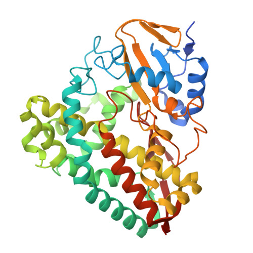
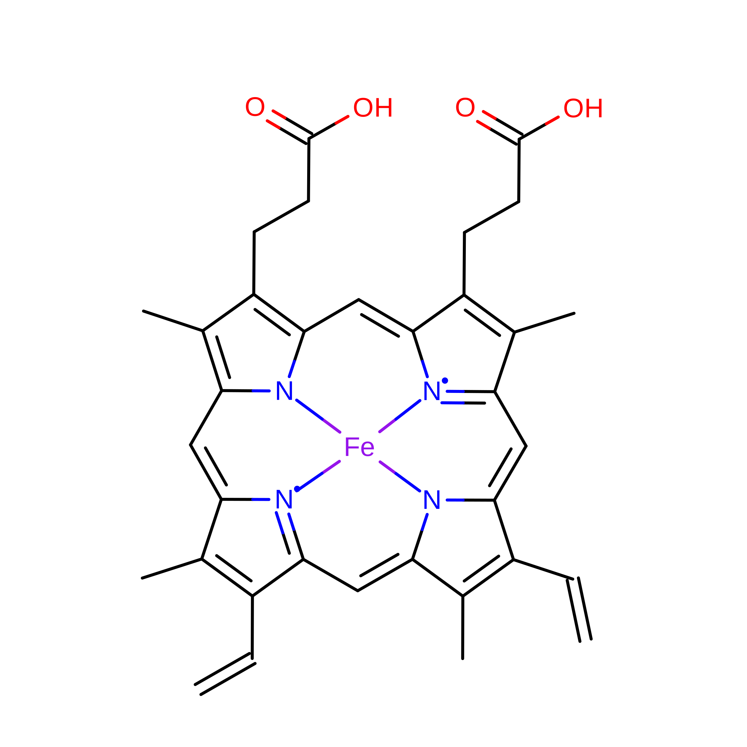
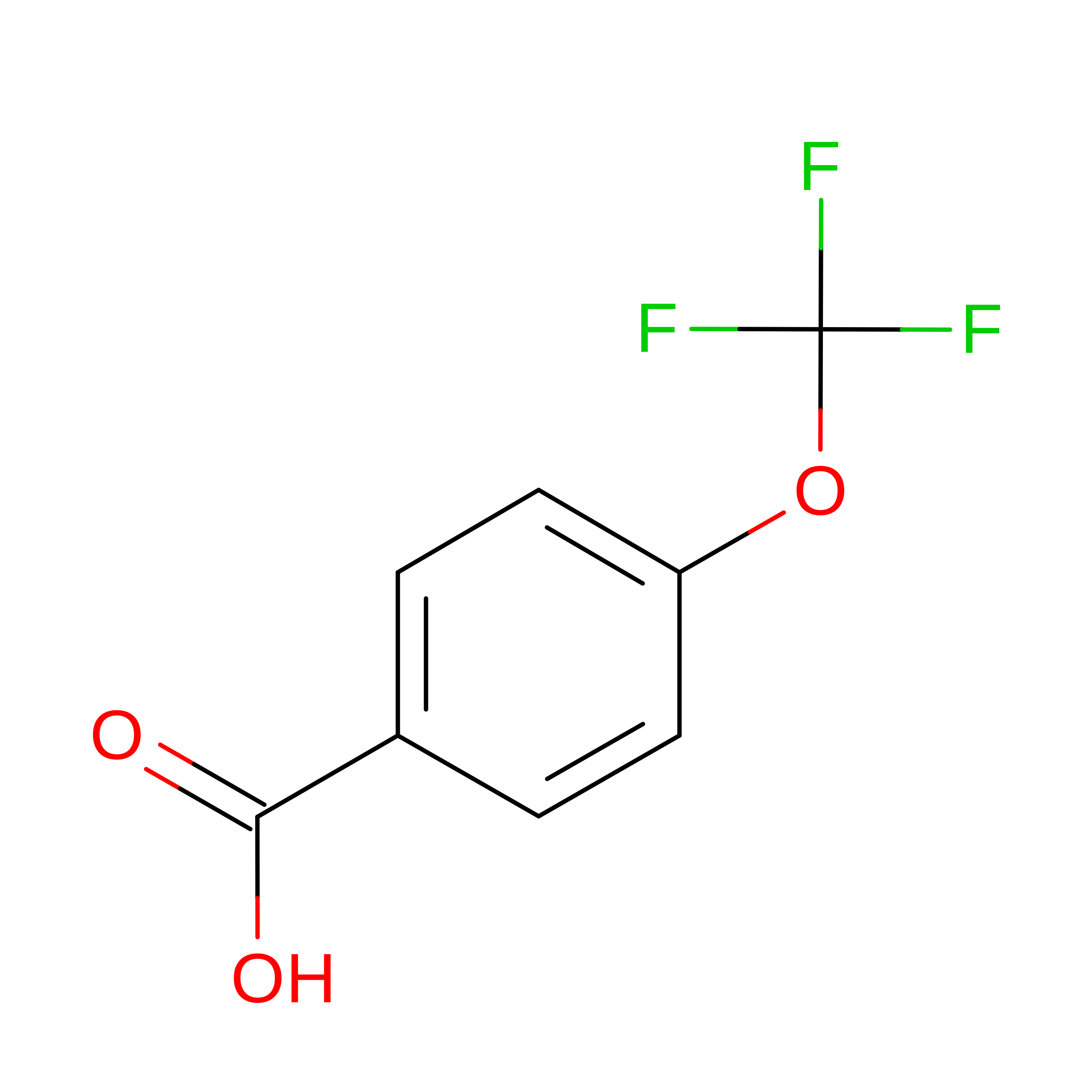
Crystal structure of T252E-CYP199A4 in complex with 4-(Trifluoromethoxy)benzoic acid


Experimental Data Snapshot

  • Method: X-RAY DIFFRACTION
  • Resolution: 1.95 Å
  • R-Value Free: 
    0.234 (Depositor), 0.235 (DCC) 
  • R-Value Work: 
    0.188 (Depositor), 0.188 (DCC) 
  • R-Value Observed: 
    0.192 (Depositor) 

Starting Model: experimental
View more details

wwPDB Validation 3D Report Full Report

Validation slider image for 8D1C

Ligand Structure Quality Assessment 


This is version 1.1 of the entry. See complete history

Literature

Selective Oxidations Using a Cytochrome P450 Enzyme Variant Driven with Surrogate Oxygen Donors and Light.

Lee, J.H.Z.Podgorski, M.N.Moir, M.Gee, A.R.Bell, S.G.

(2022) Chemistry 28: e202201366-e202201366

  • DOI: https://doi.org/10.1002/chem.202201366
  • Primary Citation Related Structures: 
    8D1C

  • PubMed Abstract: 

    Cytochrome P450 monooxygenase enzymes are versatile catalysts, which have been adapted for multiple applications in chemical synthesis. Mutation of a highly conserved active site threonine to a glutamate can convert these enzymes into peroxygenases that utilise hydrogen peroxide (H 2 O 2 ). Here, we use the T252E-CYP199A4 variant to study peroxide-driven oxidation activity by using H 2 O 2 and urea-hydrogen peroxide (UHP). We demonstrate that the T252E variant has a higher stability to H 2 O 2 in the presence of substrate that can undergo carbon-hydrogen abstraction. This peroxygenase variant could efficiently catalyse O-demethylation and an enantioselective epoxidation reaction (94 % ee). Neither the monooxygenase nor peroxygenase pathways of the P450 demonstrated a significant kinetic isotope effect (KIE) for the oxidation of deuterated substrates. These new peroxygenase variants offer the possibility of simpler cytochrome P450 systems for selective oxidations. To demonstrate this, a light driven H 2 O 2 generating system was used to support efficient product formation with this peroxygenase enzyme.


  • Organizational Affiliation
    • Department of Chemistry, University of Adelaide, Adelaide, SA 5005, Australia.

Macromolecule Content 

  • Total Structure Weight: 45.47 kDa 
  • Atom Count: 3,370 
  • Modeled Residue Count: 393 
  • Deposited Residue Count: 410 
  • Unique protein chains: 1

Macromolecules

Find similar proteins by:|  3D Structure
Entity ID: 1
MoleculeChains  Sequence LengthOrganismDetailsImage
Cytochrome P450410Rhodopseudomonas palustris HaA2Mutation(s): 1 
Gene Names: RPB_3613
UniProt
Find proteins for Q2IU02 (Rhodopseudomonas palustris (strain HaA2))
Explore Q2IU02 
Go to UniProtKB:  Q2IU02
Entity Groups
Sequence Clusters30% Identity50% Identity70% Identity90% Identity95% Identity100% Identity
UniProt GroupQ2IU02
Sequence Annotations
Expand
Reference Sequence

Experimental Data & Validation

Experimental Data

  • Method: X-RAY DIFFRACTION
  • Resolution: 1.95 Å
  • R-Value Free:  0.234 (Depositor), 0.235 (DCC) 
  • R-Value Work:  0.188 (Depositor), 0.188 (DCC) 
  • R-Value Observed: 0.192 (Depositor) 
Space Group: P 1 21 1
Unit Cell:
Length ( Å )Angle ( ˚ )
a = 41.082α = 90
b = 51.495β = 92.25
c = 79.624γ = 90
Software Package:
Software NamePurpose
PHENIXrefinement
Aimlessdata scaling
PDB_EXTRACTdata extraction
MOSFLMdata reduction
PHASERphasing

Structure Validation

View Full Validation Report


Ligand Structure Quality Assessment 


Entry History 

& Funding Information

Deposition Data


Funding OrganizationLocationGrant Number
Australian Research Council (ARC)AustraliaDP140103229

Revision History  (Full details and data files)

  • Version 1.0: 2023-03-01
    Type: Initial release
  • Version 1.1: 2023-10-25
    Changes: Data collection, Refinement description