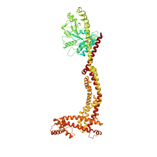
Structural mechanism of mitochondrial membrane remodelling by human OPA1.
von der Malsburg, A., Sapp, G.M., Zuccaro, K.E., von Appen, A., Moss 3rd, F.R., Kalia, R., Bennett, J.A., Abriata, L.A., Dal Peraro, M., van der Laan, M., Frost, A., Aydin, H.(2023) Nature 620: 1101-1108
- PubMed: 37612504 Search on PubMedSearch on PubMed Central
- DOI: https://doi.org/10.1038/s41586-023-06441-6
- Primary Citation Related Structures:
8CT1, 8CT9 - PubMed Abstract:
Distinct morphologies of the mitochondrial network support divergent metabolic and regulatory processes that determine cell function and fate 1-3 . The mechanochemical GTPase optic atrophy 1 (OPA1) influences the architecture of cristae and catalyses the fusion of the mitochondrial inner membrane 4,5 . Despite its fundamental importance, the molecular mechanisms by which OPA1 modulates mitochondrial morphology are unclear. Here, using a combination of cellular and structural analyses, we illuminate the molecular mechanisms that are key to OPA1-dependent membrane remodelling and fusion. Human OPA1 embeds itself into cardiolipin-containing membranes through a lipid-binding paddle domain. A conserved loop within the paddle domain inserts deeply into the bilayer, further stabilizing the interactions with cardiolipin-enriched membranes. OPA1 dimerization through the paddle domain promotes the helical assembly of a flexible OPA1 lattice on the membrane, which drives mitochondrial fusion in cells. Moreover, the membrane-bending OPA1 oligomer undergoes conformational changes that pull the membrane-inserting loop out of the outer leaflet and contribute to the mechanics of membrane remodelling. Our findings provide a structural framework for understanding how human OPA1 shapes mitochondrial morphology and show us how human disease mutations compromise OPA1 functions.
- Medical Biochemistry & Molecular Biology, Center for Molecular Signaling, PZMS, Saarland University Medical School, Homburg, Germany.
Organizational Affiliation: