8CSH

Structure of the DNA binding domain of pSK1 Par partition protein bound to centromere DNA


Experimental Data Snapshot

  • Method: X-RAY DIFFRACTION
  • Resolution: 2.25 Å
  • R-Value Free: 0.249 
  • R-Value Work: 0.202 
  • R-Value Observed: 0.207 

wwPDB Validation   3D Report Full Report


This is version 1.2 of the entry. See complete history


Literature

Molecular Analysis of pSK1 par: A Novel Plasmid Partitioning System Encoded by Staphylococcal Multiresistance Plasmids.

Chan, H.Y.Jensen, S.O.LeBard, R.J.Figgett, W.A.Lai, E.Simpson, A.E.Brzoska, A.J.Davies, D.S.Connolly, A.M.Cordwell, S.J.Travis, B.A.Salinas, R.Skurray, R.A.Firth, N.Schumacher, M.A.

(2022) J Mol Biol 434: 167770-167770

  • DOI: https://doi.org/10.1016/j.jmb.2022.167770
  • Primary Citation of Related Structures:  
    8CSH

  • PubMed Abstract: 

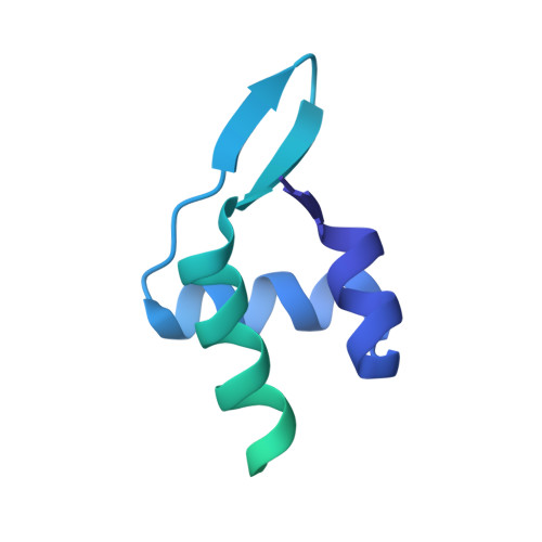
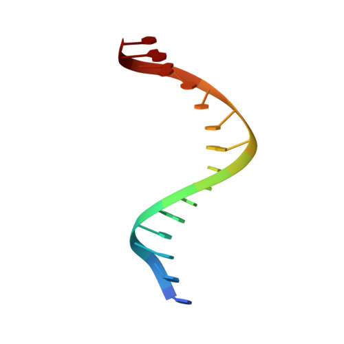
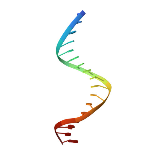
    The segregation of prokaryotic plasmids typically requires a centromere-like site and two proteins, a centromere-binding protein (CBP) and an NTPase. By contrast, a single 245 residue Par protein mediates partition of the prototypical staphylococcal multiresistance plasmid pSK1 in the absence of an identifiable NTPase component. To gain insight into centromere binding by pSK1 Par and its segregation function we performed structural, biochemical and in vivo studies. Here we show that pSK1 Par binds a centromere consisting of seven repeat elements. We demonstrate this Par-centromere interaction also mediates Par autoregulation. To elucidate the Par centromere binding mechanism, we obtained a structure of the Par N-terminal DNA-binding domain bound to centromere DNA to 2.25 Å. The pSK1 Par structure, which harbors a winged-helix-turn-helix (wHTH), is distinct from other plasmid CBP structures but shows homology to the B. subtilis chromosome segregation protein, RacA. Biochemical studies suggest the region C-terminal to the Par wHTH forms coiled coils and mediates oligomerization. Fluorescence microscopy analyses show that pSK1 Par enhances the separation of plasmids from clusters, driving effective segregation upon cell division. Combined the data provide insight into the molecular properties of a single protein partition system.


  • Organizational Affiliation

    School of Life and Environmental Sciences, University of Sydney, New South Wales 2006, Australia.


Macromolecules

Find similar proteins by:  (by identity cutoff)  |  3D Structure
Entity ID: 2
MoleculeChains Sequence LengthOrganismDetailsImage
ParB [auth D],
C [auth A]
170unidentified plasmidMutation(s): 0 
Gene Names: par
UniProt
Find proteins for Q9L8I7 (Staphylococcus aureus)
Explore Q9L8I7 
Go to UniProtKB:  Q9L8I7
Entity Groups  
Sequence Clusters30% Identity50% Identity70% Identity90% Identity95% Identity100% Identity
UniProt GroupQ9L8I7
Sequence Annotations
Expand
  • Reference Sequence

Find similar nucleic acids by:  Sequence   |   3D Structure  

Entity ID: 1
MoleculeChains LengthOrganismImage
DNA (5'-D(P*TP*GP*TP*TP*AP*GP*GP*TP*AP*CP*CP*TP*AP*AP*C)-3')A [auth T]15synthetic construct
Sequence Annotations
Expand
  • Reference Sequence

Find similar nucleic acids by:  Sequence   |   3D Structure  

Entity ID: 3
MoleculeChains LengthOrganismImage
DNA (5'-D(*TP*AP*GP*TP*TP*AP*GP*GP*TP*AP*CP*CP*TP*AP*AP*CP*A)-3')D [auth Y]17synthetic construct
Sequence Annotations
Expand
  • Reference Sequence
Experimental Data & Validation

Experimental Data

  • Method: X-RAY DIFFRACTION
  • Resolution: 2.25 Å
  • R-Value Free: 0.249 
  • R-Value Work: 0.202 
  • R-Value Observed: 0.207 
  • Space Group: C 1 2 1
Unit Cell:
Length ( Å )Angle ( ˚ )
a = 70.377α = 90
b = 91.069β = 119.85
c = 59.184γ = 90
Software Package:
Software NamePurpose
MOSFLMdata reduction
SCALAdata scaling
PHENIXrefinement
PDB_EXTRACTdata extraction
AutoSolphasing

Structure Validation

View Full Validation Report


Entry History & Funding Information

Deposition Data


Funding OrganizationLocationGrant Number
National Institutes of Health/National Institute of General Medical Sciences (NIH/NIGMS)United StatesR35-GM130290

Revision History  (Full details and data files)

  • Version 1.0: 2022-08-10
    Type: Initial release
  • Version 1.1: 2022-08-24
    Changes: Database references
  • Version 1.2: 2024-02-14
    Changes: Data collection