A multichaperone condensate enhances protein folding in the endoplasmic reticulum.
Leder, A., Mas, G., Szentgyorgyi, V., Jakob, R.P., Maier, T., Spang, A., Hiller, S.(2025) Nat Cell Biol 27: 1422-1430
- PubMed: 40789936 Search on PubMedSearch on PubMed Central
- DOI: https://doi.org/10.1038/s41556-025-01730-w
- Primary Citation Related Structures:
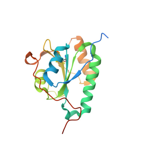
8CPQ - PubMed Abstract:
Protein folding in the endoplasmic reticulum (ER) relies on a network of molecular chaperones that facilitates the folding and maturation of client proteins. How the ER chaperones organize in a supramolecular manner to exert their cooperativity has, however, remained unclear. Here we report the discovery of a multichaperone condensate in the ER lumen, which is formed around the chaperone PDIA6 during protein folding homeostasis. The condensates form in a Ca 2+ -dependent manner and we resolve the underlying mechanism at the atomic and cellular levels. The PDIA6 condensates recruit further chaperones-Hsp70 BiP, J-domain protein ERdj3, disulfide isomerase PDIA1 and Hsp90 Grp94-which constitute some of the essential components of the early folding machinery. The chaperone condensates enhance folding of proteins, such as proinsulin, and prevent protein misfolding in the ER lumen. The PDIA6-scaffolded chaperone condensates hence provide the functional basis for spatial and temporal coordination of the dynamic ER chaperone network.
- Biozentrum, University of Basel, Basel, Switzerland.
Organizational Affiliation: