8CAT

The NADPH binding site on beef liver catalase


Experimental Data Snapshot

  • Method: X-RAY DIFFRACTION
  • Resolution: 2.50 Å
  • R-Value Work: 0.191 

wwPDB Validation   3D Report Full Report


This is version 1.5 of the entry. See complete history


Literature

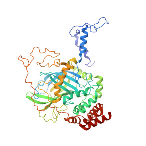
The NADPH binding site on beef liver catalase.

Fita, I.Rossmann, M.G.

(1985) Proc Natl Acad Sci U S A 82: 1604-1608

  • DOI: 10.1073/pnas.82.6.1604
  • Primary Citation of Related Structures:  
    7CAT, 8CAT

  • PubMed Abstract: 
  • Beef liver and human erythrocyte catalases (EC 1.11.1.6) bind NADP tenaciously [Kirkman, H. N. & Gaetani, G. F. (1984) Proc. Natl. Acad. Sci. USA 81, 4343-4348]. The position of NADP on beef liver catalase corresponds to the carboxyl-terminal polypeptide hinge in Penicillium vitale fungal catalase, which connects the common catalase structure to the additional flavodoxin-like domain ...

    Beef liver and human erythrocyte catalases (EC 1.11.1.6) bind NADP tenaciously [Kirkman, H. N. & Gaetani, G. F. (1984) Proc. Natl. Acad. Sci. USA 81, 4343-4348]. The position of NADP on beef liver catalase corresponds to the carboxyl-terminal polypeptide hinge in Penicillium vitale fungal catalase, which connects the common catalase structure to the additional flavodoxin-like domain. In contrast to nearly all other known structures of protein-bound NADP, NAD, and FAD, the NADP molecule of beef liver catalase is folded into a right-handed helix and bound, in part, in the vicinity of the carboxyl end of two alpha-helices. A water molecule (W7) occupies a pseudosubstrate site close to the C4 position of the nicotinamide and is hydrogen bonded to His-304. Although the NADP and heme groups approach each other to within 13.7 A, there is no direct interaction. The function of the NADP remains a mystery.


    Related Citations: 
    • The Refined Structure of Beef Liver Catalase at 2.5 Angstroms Resolution
      Fita, I., Silva, A.M., Murthy, M.R.N., Rossmann, M.G.
      (1986) Acta Crystallogr B 42: 497
    • Comparison of Beef Liver and Penicillium Vitale Catalases
      Melik-Adamyan, W.R., Barynin, V.V., Vagin, A.A., Borisov, V.V., Vainshtein, B.K., Fita, I., Murthy, M.R.N., Rossmann, M.G.
      (1986) J Mol Biol 188: 63
    • The Active Center of Catalase
      Fita, I., Rossmann, M.G.
      (1985) J Mol Biol 185: 21
    • Structure of Beef Liver Catalase
      Murthy, M.R.N., Reid III, T.J., Sicignano, A., Tanaka, N., Rossmann, M.G.
      (1981) J Mol Biol 152: 465
    • The Structure of Beef Liver Catalase
      Murthy, M.R.N., Reid III, T.J., Sicignano, A., Tanaka, N., Rossmann, M.G.
      (1981) Кристаллография 26: 1017
    • The Structure of Beef Liver Catalase
      Murthy, M.R.N., Reid III, T.J., Sicignano, A., Tanaka, N., Rossmann, M.G.
      (1981) Sov Phys Crystallogr (engl Transl ) 26: 577
    • Structure and Heme Environment of Beef Liver Catalase at 2.5 Angstroms Resolution
      Reid III, T.J., Murthy, M.R.N., Sicignano, A., Tanaka, N., Musick, W.D.L., Rossmann, M.G.
      (1981) Proc Natl Acad Sci U S A 78: 4767
    • Crystalline Bovine Liver Catalase
      Eventoff, W., Tanaka, N., Rossmann, M.G.
      (1976) J Mol Biol 103: 799
    • The Molecular Symmetry of Bovine Liver Catalase
      Eventoff, W., Gurskaya, G.V.
      (1975) J Mol Biol 93: 55

    Organizational Affiliation

    Novira Therapeutics, a Janssen Pharmaceuticals Company, 1400 McKean Road, Spring House, PA 19477, United States.


Macromolecules
Find similar proteins by:  (by identity cutoff)  |  3D Structure
Entity ID: 1
MoleculeChainsSequence LengthOrganismDetailsImage
CATALASEA, B506Bos taurusMutation(s): 0 
Gene Names: CAT
EC: 1.11.1.6
UniProt
Find proteins for P00432 (Bos taurus)
Explore P00432 
Go to UniProtKB:  P00432
Entity Groups  
Sequence Clusters30% Identity50% Identity70% Identity90% Identity95% Identity100% Identity
UniProt GroupP00432
Protein Feature View
Expand
  • Reference Sequence
Small Molecules
Ligands 2 Unique
IDChainsName / Formula / InChI Key2D Diagram3D Interactions
NDP
Query on NDP

Download Ideal Coordinates CCD File 
D [auth A],
F [auth B]
NADPH DIHYDRO-NICOTINAMIDE-ADENINE-DINUCLEOTIDE PHOSPHATE
C21 H30 N7 O17 P3
ACFIXJIJDZMPPO-NNYOXOHSSA-N
 Ligand Interaction
HEM
Query on HEM

Download Ideal Coordinates CCD File 
C [auth A],
E [auth B]
PROTOPORPHYRIN IX CONTAINING FE
C34 H32 Fe N4 O4
KABFMIBPWCXCRK-RGGAHWMASA-L
 Ligand Interaction
Binding Affinity Annotations 
IDSourceBinding Affinity
NDP Binding MOAD:  8CAT Kd: 10 (nM) from 1 assay(s)
Experimental Data & Validation

Experimental Data

  • Method: X-RAY DIFFRACTION
  • Resolution: 2.50 Å
  • R-Value Work: 0.191 
  • Space Group: P 32 2 1
Unit Cell:
Length ( Å )Angle ( ˚ )
a = 142α = 90
b = 142β = 90
c = 103.7γ = 120

Structure Validation

View Full Validation Report


Entry History 

Deposition Data

Revision History  (Full details and data files)

  • Version 1.0: 1985-04-01
    Type: Initial release
  • Version 1.1: 2008-03-03
    Changes: Version format compliance
  • Version 1.2: 2011-07-13
    Changes: Advisory, Derived calculations, Version format compliance
  • Version 1.3: 2011-10-05
    Changes: Derived calculations
  • Version 1.4: 2012-11-21
    Changes: Derived calculations
  • Version 1.5: 2017-11-01
    Changes: Derived calculations, Other