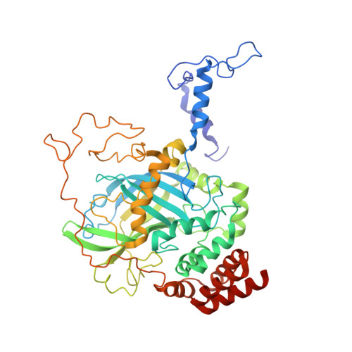
The NADPH binding site on beef liver catalase.
Fita, I., Rossmann, M.G.(1985) Proc Natl Acad Sci U S A 82: 1604-1608
- PubMed: 3856839
- DOI: https://doi.org/10.1073/pnas.82.6.1604
- Primary Citation of Related Structures:
7CAT, 8CAT - PubMed Abstract:
Beef liver and human erythrocyte catalases (EC 1.11.1.6) bind NADP tenaciously [Kirkman, H. N. & Gaetani, G. F. (1984) Proc. Natl. Acad. Sci. USA 81, 4343-4348]. The position of NADP on beef liver catalase corresponds to the carboxyl-terminal polypeptide hinge in Penicillium vitale fungal catalase, which connects the common catalase structure to the additional flavodoxin-like domain. In contrast to nearly all other known structures of protein-bound NADP, NAD, and FAD, the NADP molecule of beef liver catalase is folded into a right-handed helix and bound, in part, in the vicinity of the carboxyl end of two alpha-helices. A water molecule (W7) occupies a pseudosubstrate site close to the C4 position of the nicotinamide and is hydrogen bonded to His-304. Although the NADP and heme groups approach each other to within 13.7 A, there is no direct interaction. The function of the NADP remains a mystery.


