Understanding the Catalytic Efficiency of Two Polyester Degrading Enzymes: An Experimental and Theoretical Investigation.
Clark, M., Tornesakis, K., Konig, G., Zahn, M., Lichtenstein, B.R., Pickford, A.R., Cox, P.A.(2024) ACS Omega 9: 44724-44733
- PubMed: 39524671 Search on PubMedSearch on PubMed Central
- DOI: https://doi.org/10.1021/acsomega.4c06528
- Primary Citation Related Structures:
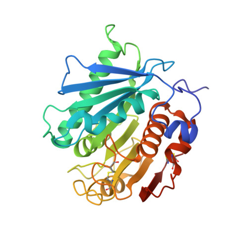
8C65 - PubMed Abstract:
The discovery of novel plastic degrading enzymes commonly relies on comparing features of the primary sequence to those of known plastic degrading enzymes. However, this approach cannot always guarantee success. This is exemplified by the different degradation rates of the two polymers poly(ethylene terephthalate) (PET) and polybutylene succinate (PBS) by two hydrolases: Is PETase from Ideonella sakaiensis and Ad Cut from Acidovorax delafieldii . Despite the enzymes showing a very high sequence identity of 82%, Is PETase shows significant hydrolysis activity for both polymers, whereas Ad Cut only shows significant hydrolysis activity for PBS. By solving the structure of Ad Cut using X-ray crystallography, and using this as the basis for computer simulations, comparisons are made between the differences in the calculated binding geometries and the catalytic results obtained from biochemical experiments. The results reveal that the low activity of Ad Cut toward PET can be explained by the low sampling of the productive conformation observed in the simulations. While the active site serine in Is PETase can closely encounter the PET carbonyl carbon, in Ad Cut it cannot: a feature that can be attributed to the shape of the catalytic binding pocket. These results yield an important insight into the design requirements for novel plastic degrading enzymes, as well as showing that computational methods can be used as a valuable tool in understanding the molecular basis for different hydrolysis activities in homologous polyesterase enzymes.
- Centre for Enzyme Innovation, University of Portsmouth, St Michael's Building, Portsmouth PO1 2DT, U.K.
Organizational Affiliation: