8C5V

Chemotaxis core signalling unit from E protein lysed E. coli cells

  • Classification: MEMBRANE PROTEIN
  • Organism(s): Escherichia coli
  • Mutation(s): No 
  • Membrane Protein: Yes  PDBTMMemProtMD

  • Deposited: 2023-01-10 Released: 2023-09-13 
  • Deposition Author(s): Cassidy, C.K., Qin, Z., Zhang, P.
  • Funding Organization(s): Wellcome Trust, Biotechnology and Biological Sciences Research Council (BBSRC), National Institutes of Health/National Institute Of Allergy and Infectious Diseases (NIH/NIAID), European Research Council (ERC)

Experimental Data Snapshot

  • Method: ELECTRON MICROSCOPY
  • Resolution: 12.0 Å
  • Aggregation State: CELL 
  • Reconstruction Method: SUBTOMOGRAM AVERAGING 

wwPDB Validation   3D Report Full Report


This is version 1.3 of the entry. See complete history


Literature

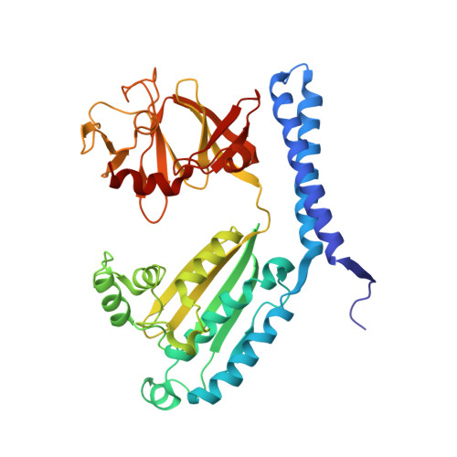
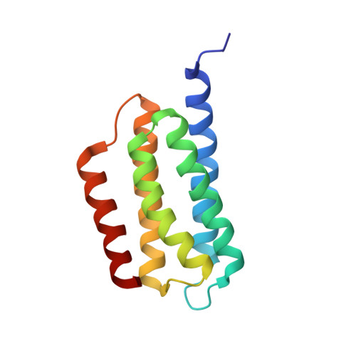
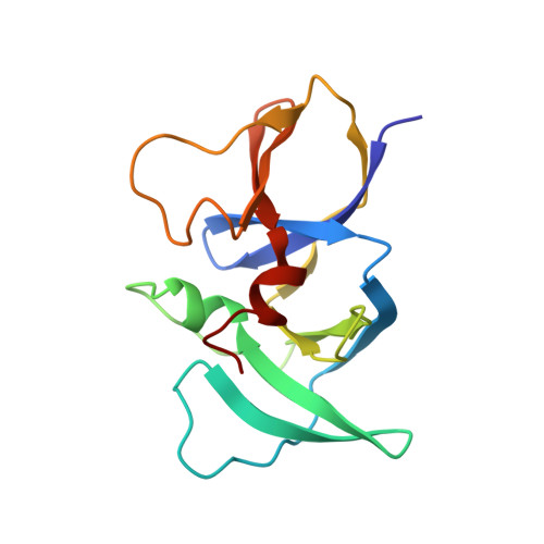
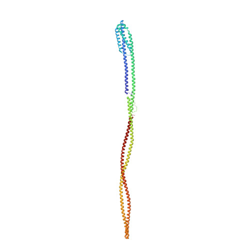
Structure of the native chemotaxis core signaling unit from phage E-protein lysed E. coli cells.

Cassidy, C.K.Qin, Z.Frosio, T.Gosink, K.Yang, Z.Sansom, M.S.P.Stansfeld, P.J.Parkinson, J.S.Zhang, P.

(2023) mBio 14: e0079323-e0079323

  • DOI: https://doi.org/10.1128/mbio.00793-23
  • Primary Citation of Related Structures:  
    8C5V

  • PubMed Abstract: 

    Bacterial chemotaxis is a ubiquitous behavior that enables cell movement toward or away from specific chemicals. It serves as an important model for understanding cell sensory signal transduction and motility. Characterization of the molecular mechanisms underlying chemotaxis is of fundamental interest and requires a high-resolution structural picture of the sensing machinery, the chemosensory array. In this study, we combine cryo-electron tomography and molecular simulation to present the complete structure of the core signaling unit, the basic building block of chemosensory arrays, from Escherichia coli . Our results provide new insight into previously poorly-resolved regions of the complex and offer a structural basis for designing new experiments to test mechanistic hypotheses.


  • Organizational Affiliation

    Diamond Light Source , Didcot, United Kingdom.


Macromolecules
Find similar proteins by:  (by identity cutoff)  |  3D Structure
Entity ID: 1
MoleculeChains Sequence LengthOrganismDetailsImage
Chemotaxis protein CheA
A, B
391Escherichia coliMutation(s): 0 
EC: 2.7.13.3
UniProt
Find proteins for P07363 (Escherichia coli (strain K12))
Explore P07363 
Go to UniProtKB:  P07363
Entity Groups  
Sequence Clusters30% Identity50% Identity70% Identity90% Identity95% Identity100% Identity
UniProt GroupP07363
Sequence Annotations
Expand
  • Reference Sequence
Find similar proteins by:  (by identity cutoff)  |  3D Structure
Entity ID: 2
MoleculeChains Sequence LengthOrganismDetailsImage
Chemotaxis protein CheA
C, D
131Escherichia coliMutation(s): 0 
EC: 2.7.13.3
UniProt
Find proteins for P07363 (Escherichia coli (strain K12))
Explore P07363 
Go to UniProtKB:  P07363
Entity Groups  
Sequence Clusters30% Identity50% Identity70% Identity90% Identity95% Identity100% Identity
UniProt GroupP07363
Sequence Annotations
Expand
  • Reference Sequence
Find similar proteins by:  (by identity cutoff)  |  3D Structure
Entity ID: 3
MoleculeChains Sequence LengthOrganismDetailsImage
Chemotaxis protein CheW
E, F, G, H
143Escherichia coliMutation(s): 0 
UniProt
Find proteins for P0A964 (Escherichia coli (strain K12))
Explore P0A964 
Go to UniProtKB:  P0A964
Entity Groups  
Sequence Clusters30% Identity50% Identity70% Identity90% Identity95% Identity100% Identity
UniProt GroupP0A964
Sequence Annotations
Expand
  • Reference Sequence
Find similar proteins by:  (by identity cutoff)  |  3D Structure
Entity ID: 4
MoleculeChains Sequence LengthOrganismDetailsImage
Methyl-accepting chemotaxis protein I
I, J, K, L, M
I, J, K, L, M, N, O, P, Q, R, S, T
516Escherichia coliMutation(s): 0 
Membrane Entity: Yes 
UniProt
Find proteins for P02942 (Escherichia coli (strain K12))
Explore P02942 
Go to UniProtKB:  P02942
Entity Groups  
Sequence Clusters30% Identity50% Identity70% Identity90% Identity95% Identity100% Identity
UniProt GroupP02942
Sequence Annotations
Expand
  • Reference Sequence
Experimental Data & Validation

Experimental Data

  • Method: ELECTRON MICROSCOPY
  • Resolution: 12.0 Å
  • Aggregation State: CELL 
  • Reconstruction Method: SUBTOMOGRAM AVERAGING 
EM Software:
TaskSoftware PackageVersion
MODEL REFINEMENTISOLDE1.5

Structure Validation

View Full Validation Report


Entry History & Funding Information

Deposition Data


Funding OrganizationLocationGrant Number
Wellcome TrustUnited Kingdom203141/Z/16/Z
Biotechnology and Biological Sciences Research Council (BBSRC)United KingdomBBSRC: B5R00550 B500.01
National Institutes of Health/National Institute Of Allergy and Infectious Diseases (NIH/NIAID)United StatesP50AI150481
Wellcome TrustUnited Kingdom206422/Z/17/Z
Biotechnology and Biological Sciences Research Council (BBSRC)United KingdomBB/S003339/1
European Research Council (ERC)European Union101021133

Revision History  (Full details and data files)

  • Version 1.0: 2023-09-13
    Type: Initial release
  • Version 1.1: 2023-09-20
    Changes: Structure summary
  • Version 1.2: 2023-10-11
    Changes: Database references
  • Version 1.3: 2023-11-29
    Changes: Database references