8C3F | pdb_00008c3f

Double mutant I(L177)H/F(M197)H structure of Photosynthetic Reaction Center From Cereibacter sphaeroides strain RV


Experimental Data Snapshot

  • Method: X-RAY DIFFRACTION
  • Resolution: 2.60 Å
  • R-Value Free: 
    0.220 (Depositor), 0.194 (DCC) 
  • R-Value Work: 
    0.194 (Depositor) 
  • R-Value Observed: 
    0.196 (Depositor) 

Starting Model: experimental
View more details

wwPDB Validation 3D Report Full Report

Validation slider image for 8C3F

Ligand Structure Quality Assessment 


This is version 1.1 of the entry. See complete history

Literature

Properties and Crystal Structure of the Cereibacter sphaeroides Photosynthetic Reaction Center with Double Amino Acid Substitution I(L177)H + F(M197)H.

Fufina, T.Y.Selikhanov, G.K.Gabdulkhakov, A.G.Vasilieva, L.G.

(2023) Membranes (Basel) 13

  • DOI: https://doi.org/10.3390/membranes13020157
  • Primary Citation Related Structures: 
    8C3F

  • PubMed Abstract: 

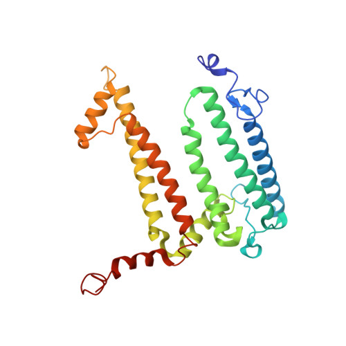
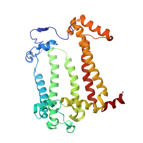
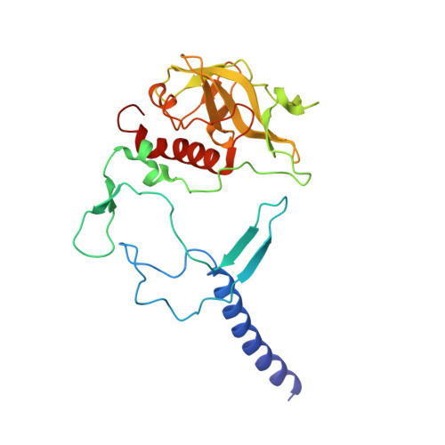
    The photosynthetic reaction center of the purple bacterium Cereibacter sphaeroides with two site-directed mutations Ile-L177-His and M197 Phe-His is of double interest. The substitution I(L177)H results in strong binding of a bacteriochlorophyll molecule with L-subunit. The second mutation F(M197)H introduces a new H-bond between the C2-acetyl carbonyl group of the bacteriochlorophyll P B and His-M197, which is known to enhance the stability of the complex. Due to this H-bond, π -electron system of P finds itself connected to an extensive H-bonding network on the periplasmic surface of the complex. The crystal structure of the double mutant reaction center obtained with 2.6 Å resolution allows clarifying consequences of the Ile L177 - His substitution. The value of the P/P + midpoint potential in the double mutant RC was found to be ~20 mV less than the sum of potentials measured in the two RCs with single mutations I(L177)H and F(M197)H. The protein environment of the BChls P A and B B were found to be similar to that in the RC with single substitution I(L177)H, whereas an altered pattern of the H-bonding networks was found in the vicinity of bacteriochlorophyll P B . The data obtained are consistent with our previous assumption on a correlation between the bulk of the H-bonding network connected with the π-electron system of the primary electron donor P and the value of its oxidation potential.


  • Organizational Affiliation
    • Federal Research Center Pushchino Scientific Center for Biological Research PSCBR, Institute of Basic Biological Problems, Russian Academy of Sciences, Institutskaya Street 2, 142290 Pushchino, Russia.

Macromolecule Content 

  • Total Structure Weight: 104.12 kDa 
  • Atom Count: 7,413 
  • Modeled Residue Count: 822 
  • Deposited Residue Count: 825 
  • Unique protein chains: 3

Macromolecules

Find similar proteins by:|  3D Structure
Entity ID: 1
MoleculeChains  Sequence LengthOrganismDetailsImage
Reaction center protein L chainA [auth L]281Cereibacter sphaeroides 2.4.1Mutation(s): 2 
Gene Names: pufL
Membrane Entity: Yes 
UniProt
Find proteins for Q3J1A5 (Cereibacter sphaeroides (strain ATCC 17023 / DSM 158 / JCM 6121 / CCUG 31486 / LMG 2827 / NBRC 12203 / NCIMB 8253 / ATH 2.4.1.))
Explore Q3J1A5 
Go to UniProtKB:  Q3J1A5
Entity Groups
Sequence Clusters30% Identity50% Identity70% Identity90% Identity95% Identity100% Identity
UniProt GroupQ3J1A5
Sequence Annotations
Expand
Reference Sequence
Find similar proteins by:|  3D Structure
Entity ID: 2
MoleculeChains  Sequence LengthOrganismDetailsImage
Reaction center protein M chainB [auth M]303Cereibacter sphaeroides 2.4.1Mutation(s): 2 
Gene Names: pufM
Membrane Entity: Yes 
UniProt
Find proteins for Q3J1A6 (Cereibacter sphaeroides (strain ATCC 17023 / DSM 158 / JCM 6121 / CCUG 31486 / LMG 2827 / NBRC 12203 / NCIMB 8253 / ATH 2.4.1.))
Explore Q3J1A6 
Go to UniProtKB:  Q3J1A6
Entity Groups
Sequence Clusters30% Identity50% Identity70% Identity90% Identity95% Identity100% Identity
UniProt GroupQ3J1A6
Sequence Annotations
Expand
Reference Sequence
Find similar proteins by:|  3D Structure
Entity ID: 3
MoleculeChains  Sequence LengthOrganismDetailsImage
Reaction center protein H chainC [auth H]241Cereibacter sphaeroides 2.4.1Mutation(s): 0 
Gene Names: puhA
Membrane Entity: Yes 
UniProt
Find proteins for Q3J170 (Cereibacter sphaeroides (strain ATCC 17023 / DSM 158 / JCM 6121 / CCUG 31486 / LMG 2827 / NBRC 12203 / NCIMB 8253 / ATH 2.4.1.))
Explore Q3J170 
Go to UniProtKB:  Q3J170
Entity Groups
Sequence Clusters30% Identity50% Identity70% Identity90% Identity95% Identity100% Identity
UniProt GroupQ3J170
Sequence Annotations
Expand
Reference Sequence

Small Molecules

Ligands 15 Unique
IDChains Name / Formula / InChI Key2D Diagram3D Interactions
CDL
(Subject of Investigation/LOI)

Query on CDL


Download:Ideal Coordinates CCD File
DA [auth M]CARDIOLIPIN
C81 H156 O17 P2
XVTUQDWPJJBEHJ-KZCWQMDCSA-L
BCL
(Subject of Investigation/LOI)

Query on BCL


Download:Ideal Coordinates CCD File
H [auth L],
P [auth L],
S [auth M],
V [auth M]
BACTERIOCHLOROPHYLL A
C55 H74 Mg N4 O6
DSJXIQQMORJERS-AGGZHOMASA-M
BPB
(Subject of Investigation/LOI)

Query on BPB


Download:Ideal Coordinates CCD File
I [auth L],
W [auth M]
BACTERIOPHEOPHYTIN B
C55 H74 N4 O6
SFKCKJXMIAKQMY-GTTFDWDMSA-N
U10
(Subject of Investigation/LOI)

Query on U10


Download:Ideal Coordinates CCD File
J [auth L],
Y [auth M]
UBIQUINONE-10
C59 H90 O4
ACTIUHUUMQJHFO-UPTCCGCDSA-N
SPN
(Subject of Investigation/LOI)

Query on SPN


Download:Ideal Coordinates CCD File
Z [auth M]SPEROIDENONE
C41 H70 O2
GWQAMGYOEYXWJF-YCDPMLDASA-N
LDA

Query on LDA


Download:Ideal Coordinates CCD File
EA [auth M]
FA [auth M]
GA [auth M]
HA [auth M]
MA [auth H]
EA [auth M],
FA [auth M],
GA [auth M],
HA [auth M],
MA [auth H],
N [auth L],
O [auth L],
T [auth M]
LAURYL DIMETHYLAMINE-N-OXIDE
C14 H31 N O
SYELZBGXAIXKHU-UHFFFAOYSA-N
HTO

Query on HTO


Download:Ideal Coordinates CCD File
G [auth L],
R [auth M]
HEPTANE-1,2,3-TRIOL
C7 H16 O3
HXYCHJFUBNTKQR-RNFRBKRXSA-N
TRS

Query on TRS


Download:Ideal Coordinates CCD File
LA [auth H]2-AMINO-2-HYDROXYMETHYL-PROPANE-1,3-DIOL
C4 H12 N O3
LENZDBCJOHFCAS-UHFFFAOYSA-O
PO4

Query on PO4


Download:Ideal Coordinates CCD File
CA [auth M],
K [auth L],
L
PHOSPHATE ION
O4 P
NBIIXXVUZAFLBC-UHFFFAOYSA-K
GOL

Query on GOL


Download:Ideal Coordinates CCD File
M [auth L]GLYCEROL
C3 H8 O3
PEDCQBHIVMGVHV-UHFFFAOYSA-N
DIO

Query on DIO


Download:Ideal Coordinates CCD File
F [auth L]1,4-DIETHYLENE DIOXIDE
C4 H8 O2
RYHBNJHYFVUHQT-UHFFFAOYSA-N
EDO

Query on EDO


Download:Ideal Coordinates CCD File
D [auth L]
E [auth L]
IA [auth M]
JA [auth H]
KA [auth H]
D [auth L],
E [auth L],
IA [auth M],
JA [auth H],
KA [auth H],
Q [auth M],
U [auth M]
1,2-ETHANEDIOL
C2 H6 O2
LYCAIKOWRPUZTN-UHFFFAOYSA-N
FE
(Subject of Investigation/LOI)

Query on FE


Download:Ideal Coordinates CCD File
X [auth M]FE (III) ION
Fe
VTLYFUHAOXGGBS-UHFFFAOYSA-N
K

Query on K


Download:Ideal Coordinates CCD File
AA [auth M],
NA [auth H]
POTASSIUM ION
K
NPYPAHLBTDXSSS-UHFFFAOYSA-N
CL

Query on CL


Download:Ideal Coordinates CCD File
BA [auth M]CHLORIDE ION
Cl
VEXZGXHMUGYJMC-UHFFFAOYSA-M

Experimental Data & Validation

Experimental Data

  • Method: X-RAY DIFFRACTION
  • Resolution: 2.60 Å
  • R-Value Free:  0.220 (Depositor), 0.194 (DCC) 
  • R-Value Work:  0.194 (Depositor) 
  • R-Value Observed: 0.196 (Depositor) 
Space Group: P 31 2 1
Unit Cell:
Length ( Å )Angle ( ˚ )
a = 139.346α = 90
b = 139.346β = 90
c = 184.574γ = 120
Software Package:
Software NamePurpose
REFMACrefinement
PHENIXrefinement
XDSdata reduction
XSCALEdata scaling
PHASERphasing

Structure Validation

View Full Validation Report


Ligand Structure Quality Assessment 


Entry History 

& Funding Information

Deposition Data


Funding OrganizationLocationGrant Number
Not funded--

Revision History  (Full details and data files)

  • Version 1.0: 2023-03-08
    Type: Initial release
  • Version 1.1: 2024-02-07
    Changes: Data collection