8C2E

Structure of 14-3-3 sigma delta C with electrophilic peptide 4IEA-5


Experimental Data Snapshot

  • Method: X-RAY DIFFRACTION
  • Resolution: 2.20 Å
  • R-Value Free: 0.221 
  • R-Value Work: 0.175 
  • R-Value Observed: 0.178 

wwPDB Validation   3D Report Full Report


Ligand Structure Quality Assessment 


This is version 1.1 of the entry. See complete history


Literature

A simple method for developing lysine targeted covalent protein reagents.

Gabizon, R.Tivon, B.Reddi, R.N.van den Oetelaar, M.C.M.Amartely, H.Cossar, P.J.Ottmann, C.London, N.

(2023) Nat Commun 14: 7933-7933

  • DOI: https://doi.org/10.1038/s41467-023-42632-5
  • Primary Citation of Related Structures:  
    8C2E, 8C2F

  • PubMed Abstract: 

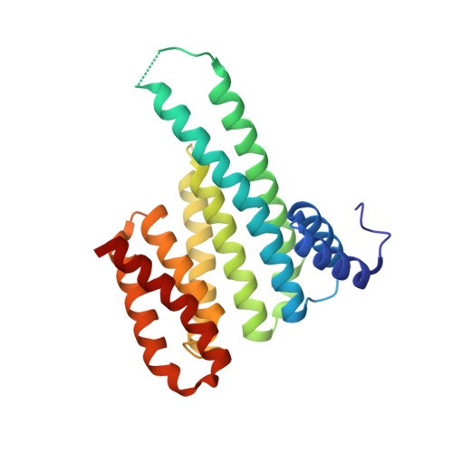
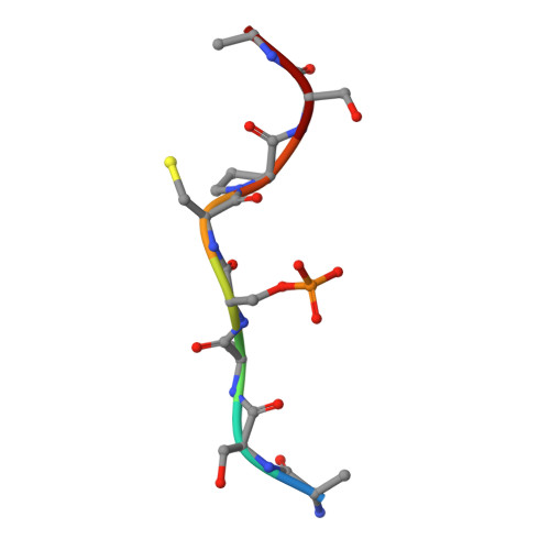
    Peptide-based covalent probes can target shallow protein surfaces not typically addressable using small molecules, yet there is a need for versatile approaches to convert native peptide sequences into covalent binders that can target a broad range of residues. Here we report protein-based thio-methacrylate esters-electrophiles that can be installed easily on unprotected peptides and proteins via cysteine side chains, and react efficiently and selectively with cysteine and lysine side chains on the target. Methacrylate phosphopeptides derived from 14-3-3-binding proteins irreversibly label 14-3-3σ via either lysine or cysteine residues, depending on the position of the electrophile. Methacrylate peptides targeting a conserved lysine residue exhibit pan-isoform binding of 14-3-3 proteins both in lysates and in extracellular media. Finally, we apply this approach to develop protein-based covalent binders. A methacrylate-modified variant of the colicin E9 immunity protein irreversibly binds to the E9 DNAse, resulting in significantly higher thermal stability relative to the non-covalent complex. Our approach offers a simple and versatile route to convert peptides and proteins into potent covalent binders.


  • Organizational Affiliation

    Department of Chemical and Structural Biology, The Weizmann Institute of Science, Rehovot, 7610001, Israel.


Macromolecules
Find similar proteins by:  (by identity cutoff)  |  3D Structure
Entity ID: 1
MoleculeChains Sequence LengthOrganismDetailsImage
14-3-3 protein sigma236Homo sapiensMutation(s): 0 
Gene Names: SFNHME1
UniProt & NIH Common Fund Data Resources
Find proteins for P31947 (Homo sapiens)
Explore P31947 
Go to UniProtKB:  P31947
PHAROS:  P31947
GTEx:  ENSG00000175793 
Entity Groups  
Sequence Clusters30% Identity50% Identity70% Identity90% Identity95% Identity100% Identity
UniProt GroupP31947
Sequence Annotations
Expand
  • Reference Sequence

Find similar proteins by:  Sequence   |   3D Structure  

Entity ID: 2
MoleculeChains Sequence LengthOrganismDetailsImage
ARG-SER-ALA-SEP-CYS-PRO-SER-LEUB [auth P]8Homo sapiensMutation(s): 0 
Sequence Annotations
Expand
  • Reference Sequence
Experimental Data & Validation

Experimental Data

  • Method: X-RAY DIFFRACTION
  • Resolution: 2.20 Å
  • R-Value Free: 0.221 
  • R-Value Work: 0.175 
  • R-Value Observed: 0.178 
  • Space Group: C 2 2 21
Unit Cell:
Length ( Å )Angle ( ˚ )
a = 82.513α = 90
b = 111.724β = 90
c = 63.077γ = 90
Software Package:
Software NamePurpose
REFMACrefinement
DIALSdata reduction
Aimlessdata scaling
MOLREPphasing
Cootmodel building

Structure Validation

View Full Validation Report


Ligand Structure Quality Assessment 


Entry History & Funding Information

Deposition Data


Funding OrganizationLocationGrant Number
Netherlands Organisation for Scientific Research (NWO)Netherlands--

Revision History  (Full details and data files)

  • Version 1.0: 2023-11-01
    Type: Initial release
  • Version 1.1: 2023-12-13
    Changes: Database references