A Combined in Silico and Structural Study Opens New Perspectives on Aliphatic Sulfonamides, a Still Poorly Investigated Class of CA Inhibitors.
Langella, E., Esposito, D., Monti, S.M., Supuran, C.T., De Simone, G., Alterio, V.(2023) Biology (Basel) 12
- PubMed: 36829558 Search on PubMedSearch on PubMed Central
- DOI: https://doi.org/10.3390/biology12020281
- Primary Citation Related Structures:
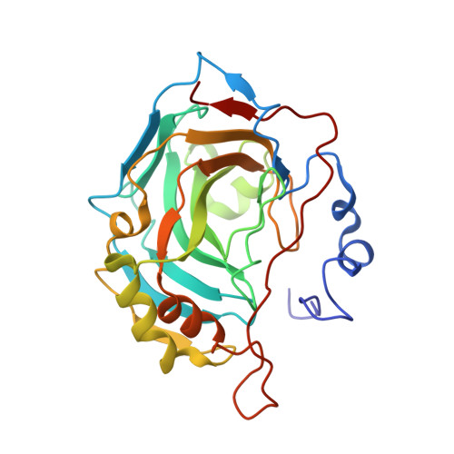
8C0Q, 8C0R - PubMed Abstract:
Aliphatic sulfonamides are an interesting class of carbonic anhydrase inhibitors (CAIs) proven to be effective for several carbonic anhydrase (CA) isoforms involved in pathologic states. Here we report the crystallographic structures of hCA II in complex with two aliphatic sulfonamides incorporating coumarin rings, which showed a good inhibition and selectivity for this isoform. Although these two molecules have a very similar chemical structure, differing only in the substitution of the two aliphatic hydrogen atoms with two fluorine atoms, they adopt a significantly different binding mode within the enzyme active site. Theoretical binding free energy calculations, performed to rationalize these data, showed that a delicate balance of electrostatic and steric effects modulate the protein-ligand interactions. Data presented here can be fruitfully used for the rational design of novel and effective isozyme-specific inhibitor molecules.
- Institute of Biostructures and Bioimaging-CNR, Via Pietro Castellino 111, 80131 Naples, Italy.
Organizational Affiliation: