Small-molecule positive allosteric modulation of homomeric kainate receptors GluK1-3: development of screening assays and insight into GluK3 structure.
Bay, Y., Venskutonyte, R., Frantsen, S.M., Thorsen, T.S., Musgaard, M., Frydenvang, K., Francotte, P., Pirotte, B., Biggin, P.C., Kristensen, A.S., Boesen, T., Pickering, D.S., Gajhede, M., Kastrup, J.S.(2024) FEBS J 291: 1506-1529
- PubMed: 38145505 Search on PubMed
- DOI: https://doi.org/10.1111/febs.17046
- Primary Citation Related Structures:
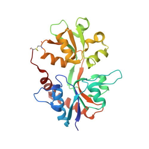
8BST, 8BSU - PubMed Abstract:
The kainate receptors GluK1-3 (glutamate receptor ionotropic, kainate receptors 1-3) belong to the family of ionotropic glutamate receptors and are essential for fast excitatory neurotransmission in the brain, and are associated with neurological and psychiatric diseases. How these receptors can be modulated by small-molecule agents is not well understood, especially for GluK3. We show that the positive allosteric modulator BPAM344 can be used to establish robust calcium-sensitive fluorescence-based assays to test agonists, antagonists, and positive allosteric modulators of GluK1-3. The half-maximal effective concentration (EC 50 ) of BPAM344 for potentiating the response of 100 μm kainate was determined to be 26.3 μm for GluK1, 75.4 μm for GluK2, and 639 μm for GluK3. Domoate was found to be a potent agonist for GluK1 and GluK2, with an EC 50 of 0.77 and 1.33 μm, respectively, upon co-application of 150 μm BPAM344. At GluK3, domoate acts as a very weak agonist or antagonist with a half-maximal inhibitory concentration (IC 50 ) of 14.5 μm, in presence of 500 μm BPAM344 and 100 μm kainate for competition binding. Using H523A-mutated GluK3, we determined the first dimeric structure of the ligand-binding domain by X-ray crystallography, allowing location of BPAM344, as well as zinc-, sodium-, and chloride-ion binding sites at the dimer interface. Molecular dynamics simulations support the stability of the ion sites as well as the involvement of Asp761, Asp790, and Glu797 in the binding of zinc ions. Using electron microscopy, we show that, in presence of glutamate and BPAM344, full-length GluK3 adopts a dimer-of-dimers arrangement.
- Department of Drug Design and Pharmacology, Faculty of Health and Medical Sciences, University of Copenhagen, Denmark.
Organizational Affiliation: