8BON | pdb_00008bon

Structure of the SARS-CoV-2 spike glycoprotein in complex with the macrocyclic peptide S1B3inL1


Experimental Data Snapshot

  • Method: ELECTRON MICROSCOPY
  • Resolution: 3.20 Å
  • Aggregation State: PARTICLE 
  • Reconstruction Method: SINGLE PARTICLE 

wwPDB Validation 3D Report Full Report

Validation slider image for 8BON

This is version 1.1 of the entry. See complete history

Literature

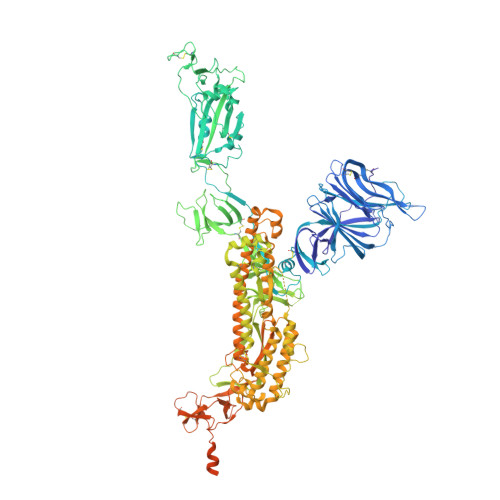
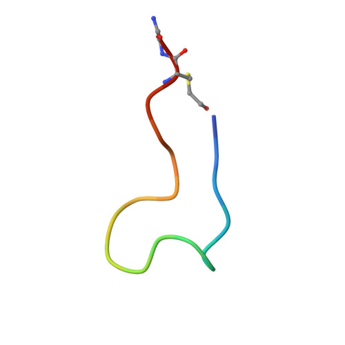
A broad-spectrum macrocyclic peptide inhibitor of the SARS-CoV-2 spike protein.

Thijssen, V.Hurdiss, D.L.Debski-Antoniak, O.J.Spence, M.A.Franck, C.Norman, A.Aggarwal, A.Mokiem, N.J.van Dongen, D.A.A.Vermeir, S.W.Liu, M.Li, W.Chatziandreou, M.Donselaar, T.Du, W.Drulyte, I.Bosch, B.J.Snijder, J.Turville, S.G.Payne, R.J.Jackson, C.J.van Kuppeveld, F.J.M.Jongkees, S.A.K.

(2023) Proc Natl Acad Sci U S A 120: e2303292120-e2303292120

  • DOI: https://doi.org/10.1073/pnas.2303292120
  • Primary Citation Related Structures: 
    8BON

  • PubMed Abstract: 

    The ongoing COVID-19 pandemic has had great societal and health consequences. Despite the availability of vaccines, infection rates remain high due to immune evasive Omicron sublineages. Broad-spectrum antivirals are needed to safeguard against emerging variants and future pandemics. We used messenger RNA (mRNA) display under a reprogrammed genetic code to find a spike-targeting macrocyclic peptide that inhibits SARS-CoV-2 (severe acute respiratory syndrome coronavirus 2) Wuhan strain infection and pseudoviruses containing spike proteins of SARS-CoV-2 variants or related sarbecoviruses. Structural and bioinformatic analyses reveal a conserved binding pocket between the receptor-binding domain, N-terminal domain, and S2 region, distal to the angiotensin-converting enzyme 2 receptor-interaction site. Our data reveal a hitherto unexplored site of vulnerability in sarbecoviruses that peptides and potentially other drug-like molecules can target.


  • Organizational Affiliation
    • Chemical Biology and Drug Discovery, Utrecht Institute for Pharmaceutical Sciences, Utrecht University, Utrecht 3584 CG, the Netherlands.

Macromolecule Content 

  • Total Structure Weight: 440.51 kDa 
  • Atom Count: 25,930 
  • Modeled Residue Count: 3,249 
  • Deposited Residue Count: 3,882 
  • Unique protein chains: 2

Macromolecules

Find similar proteins by:|  3D Structure
Entity ID: 1
MoleculeChains  Sequence LengthOrganismDetailsImage
Spike glycoprotein,FibritinA [auth B],
D [auth A],
E [auth C]
1,275Severe acute respiratory syndrome coronavirus 2Tequatrovirus T4
This entity is chimeric
Mutation(s): 9 
Gene Names: S2wac
UniProt
Find proteins for P0DTC2 (Severe acute respiratory syndrome coronavirus 2)
Explore P0DTC2 
Go to UniProtKB:  P0DTC2
Find proteins for P10104 (Enterobacteria phage T4)
Explore P10104 
Go to UniProtKB:  P10104
Entity Groups
Sequence Clusters30% Identity50% Identity70% Identity90% Identity95% Identity100% Identity
UniProt GroupsP0DTC2P10104
Glycosylation
Glycosylation Sites: 13Go to GlyGen: P0DTC2-1
Sequence Annotations
Expand
Reference Sequence
Find similar proteins by:  Sequence   |   3D Structure  
Entity ID: 2
MoleculeChains  Sequence LengthOrganismDetailsImage
Macrocyclic peptide S1B3inL1B [auth E],
C [auth D],
F
19synthetic constructMutation(s): 0 

Oligosaccharides

Help  
Entity ID: 3
MoleculeChains Length2D Diagram GlycosylationD Interactions
2-acetamido-2-deoxy-beta-D-glucopyranose-(1-4)-2-acetamido-2-deoxy-beta-D-glucopyranose
G, H, I, J, K
G, H, I, J, K, L, M, N, O, P, Q, R, S
2N-Glycosylation
Glycosylation Resources
GlyTouCan: G42666HT
GlyCosmos: G42666HT
GlyGen: G42666HT

Small Molecules

Ligands 1 Unique
IDChains Name / Formula / InChI Key2D Diagram3D Interactions
NAG

Query on NAG


Download:Ideal Coordinates CCD File
AA [auth A]
BA [auth A]
CA [auth A]
DA [auth A]
EA [auth A]
AA [auth A],
BA [auth A],
CA [auth A],
DA [auth A],
EA [auth A],
FA [auth A],
GA [auth A],
HA [auth C],
IA [auth C],
JA [auth C],
KA [auth C],
LA [auth C],
MA [auth C],
NA [auth C],
OA [auth C],
PA [auth C],
T [auth B],
U [auth B],
V [auth B],
W [auth B],
X [auth B],
Y [auth B],
Z [auth A]
2-acetamido-2-deoxy-beta-D-glucopyranose
C8 H15 N O6
OVRNDRQMDRJTHS-FMDGEEDCSA-N
Modified Residues  1 Unique
IDChains TypeFormula2D DiagramParent
CCS
Query on CCS
B [auth E],
C [auth D],
F
L-PEPTIDE LINKINGC5 H9 N O4 SCYS

Experimental Data & Validation

Experimental Data

  • Method: ELECTRON MICROSCOPY
  • Resolution: 3.20 Å
  • Aggregation State: PARTICLE 
  • Reconstruction Method: SINGLE PARTICLE 
EM Software:
TaskSoftware PackageVersion
RECONSTRUCTIONRELION3.1.3
MODEL REFINEMENTPHENIX1.19.2_4158

Structure Validation

View Full Validation Report


Entry History 

& Funding Information

Deposition Data


Funding OrganizationLocationGrant Number
Innovative Medicines InitiativeSwitzerland101005077

Revision History  (Full details and data files)

  • Version 1.0: 2023-06-28
    Type: Initial release
  • Version 1.1: 2023-07-05
    Changes: Database references