8BJJ

ExoY Nucleotidyl Cyclase domain from Vibrio nigripulchritudo MARTX toxin, bound to ATP-Mg-actin, human profilin 1 and a sulfate ion


Experimental Data Snapshot

  • Method: X-RAY DIFFRACTION
  • Resolution: 1.70 Å
  • R-Value Free: 0.195 
  • R-Value Work: 0.175 
  • R-Value Observed: 0.176 

wwPDB Validation   3D Report Full Report


Ligand Structure Quality Assessment 


This is version 1.1 of the entry. See complete history


Literature

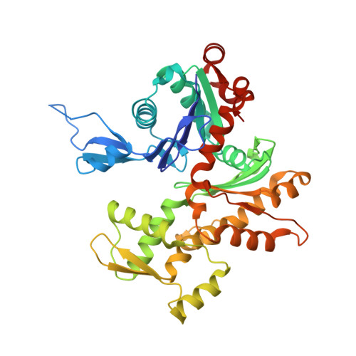
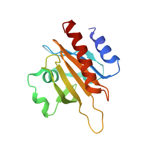
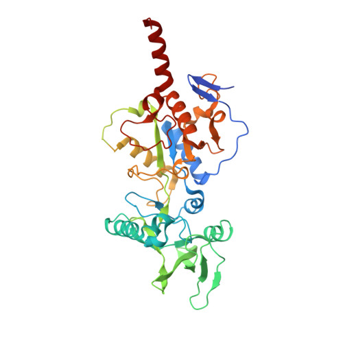
Functional and structural insights into the multi-step activation and catalytic mechanism of bacterial ExoY nucleotidyl cyclase toxins bound to actin-profilin.

Teixeira-Nunes, M.Retailleau, P.Raoux-Barbot, D.Comisso, M.Missinou, A.A.Velours, C.Plancqueel, S.Ladant, D.Mechold, U.Renault, L.

(2023) PLoS Pathog 19: e1011654-e1011654

  • DOI: https://doi.org/10.1371/journal.ppat.1011654
  • Primary Citation of Related Structures:  
    8BJH, 8BJI, 8BJJ, 8BO1, 8BR0, 8BR1

  • PubMed Abstract: 

    ExoY virulence factors are members of a family of bacterial nucleotidyl cyclases (NCs) that are activated by specific eukaryotic cofactors and overproduce cyclic purine and pyrimidine nucleotides in host cells. ExoYs act as actin-activated NC toxins. Here, we explore the Vibrio nigripulchritudo Multifunctional-Autoprocessing Repeats-in-ToXin (MARTX) ExoY effector domain (Vn-ExoY) as a model for ExoY-type members that interact with monomeric (G-actin) instead of filamentous (F-actin) actin. Vn-ExoY exhibits moderate binding affinity to free or profilin-bound G-actin but can capture the G-actin:profilin complex, preventing its spontaneous or VASP- or formin-mediated assembly at F-actin barbed ends in vitro. This mechanism may prolong the activated cofactor-bound state of Vn-ExoY at sites of active actin cytoskeleton remodelling. We present a series of high-resolution crystal structures of nucleotide-free, 3'-deoxy-ATP- or 3'-deoxy-CTP-bound Vn-ExoY, activated by free or profilin-bound G-actin-ATP/-ADP, revealing that the cofactor only partially stabilises the nucleotide-binding pocket (NBP) of NC toxins. Substrate binding induces a large, previously-unidentified, closure of their NBP, confining catalytically important residues and metal cofactors around the substrate, and facilitating the recruitment of two metal ions to tightly coordinate the triphosphate moiety of purine or pyrimidine nucleotide substrates. We validate critical residues for both the purinyl and pyrimidinyl cyclase activity of NC toxins in Vn-ExoY and its distantly-related ExoY from Pseudomonas aeruginosa, which specifically interacts with F-actin. The data conclusively demonstrate that NC toxins employ a similar two-metal-ion mechanism for catalysing the cyclisation of nucleotides of different sizes. These structural insights into the dynamics of the actin-binding interface of actin-activated ExoYs and the multi-step activation of all NC toxins offer new perspectives for the specific inhibition of class II bacterial NC enzymes.


  • Organizational Affiliation

    Université Paris-Saclay, CEA, CNRS, Institute for Integrative Biology of the Cell (I2BC), Gif-sur-Yvette, France.


Macromolecules
Find similar proteins by:  (by identity cutoff)  |  3D Structure
Entity ID: 1
MoleculeChains Sequence LengthOrganismDetailsImage
Actin, alpha skeletal muscle, intermediate form375Oryctolagus cuniculusMutation(s): 0 
UniProt
Find proteins for P68135 (Oryctolagus cuniculus)
Explore P68135 
Go to UniProtKB:  P68135
Entity Groups  
Sequence Clusters30% Identity50% Identity70% Identity90% Identity95% Identity100% Identity
UniProt GroupP68135
Sequence Annotations
Expand
  • Reference Sequence
Find similar proteins by:  (by identity cutoff)  |  3D Structure
Entity ID: 2
MoleculeChains Sequence LengthOrganismDetailsImage
Profilin-1B [auth C]139Homo sapiensMutation(s): 0 
Gene Names: PFN1
UniProt & NIH Common Fund Data Resources
Find proteins for P07737 (Homo sapiens)
Explore P07737 
Go to UniProtKB:  P07737
PHAROS:  P07737
GTEx:  ENSG00000108518 
Entity Groups  
Sequence Clusters30% Identity50% Identity70% Identity90% Identity95% Identity100% Identity
UniProt GroupP07737
Sequence Annotations
Expand
  • Reference Sequence
Find similar proteins by:  (by identity cutoff)  |  3D Structure
Entity ID: 3
MoleculeChains Sequence LengthOrganismDetailsImage
Putative Adenylate cyclaseC [auth B]413Vibrio nigripulchritudoMutation(s): 0 
EC: 4.6.1.1
UniProt
Find proteins for A0A9P1NJI6 (Vibrio nigripulchritudo)
Explore A0A9P1NJI6 
Go to UniProtKB:  A0A9P1NJI6
Entity Groups  
Sequence Clusters30% Identity50% Identity70% Identity90% Identity95% Identity100% Identity
UniProt GroupA0A9P1NJI6
Sequence Annotations
Expand
  • Reference Sequence
Small Molecules
Ligands 8 Unique
IDChains Name / Formula / InChI Key2D Diagram3D Interactions
ATP (Subject of Investigation/LOI)
Query on ATP

Download Ideal Coordinates CCD File 
D [auth A]ADENOSINE-5'-TRIPHOSPHATE
C10 H16 N5 O13 P3
ZKHQWZAMYRWXGA-KQYNXXCUSA-N
LAB (Subject of Investigation/LOI)
Query on LAB

Download Ideal Coordinates CCD File 
F [auth A]LATRUNCULIN B
C20 H29 N O5 S
NSHPHXHGRHSMIK-JRIKCGFMSA-N
PG4
Query on PG4

Download Ideal Coordinates CCD File 
O [auth B]TETRAETHYLENE GLYCOL
C8 H18 O5
UWHCKJMYHZGTIT-UHFFFAOYSA-N
TRS
Query on TRS

Download Ideal Coordinates CCD File 
G [auth A],
N [auth B]
2-AMINO-2-HYDROXYMETHYL-PROPANE-1,3-DIOL
C4 H12 N O3
LENZDBCJOHFCAS-UHFFFAOYSA-O
SO4 (Subject of Investigation/LOI)
Query on SO4

Download Ideal Coordinates CCD File 
H [auth A],
K [auth B],
L [auth B],
M [auth B]
SULFATE ION
O4 S
QAOWNCQODCNURD-UHFFFAOYSA-L
GOL
Query on GOL

Download Ideal Coordinates CCD File 
P [auth B]GLYCEROL
C3 H8 O3
PEDCQBHIVMGVHV-UHFFFAOYSA-N
PEO
Query on PEO

Download Ideal Coordinates CCD File 
I [auth C],
J [auth C]
HYDROGEN PEROXIDE
H2 O2
MHAJPDPJQMAIIY-UHFFFAOYSA-N
MG (Subject of Investigation/LOI)
Query on MG

Download Ideal Coordinates CCD File 
E [auth A]MAGNESIUM ION
Mg
JLVVSXFLKOJNIY-UHFFFAOYSA-N
Experimental Data & Validation

Experimental Data

  • Method: X-RAY DIFFRACTION
  • Resolution: 1.70 Å
  • R-Value Free: 0.195 
  • R-Value Work: 0.175 
  • R-Value Observed: 0.176 
  • Space Group: P 1 21 1
Unit Cell:
Length ( Å )Angle ( ˚ )
a = 81.186α = 90
b = 63.202β = 91.04
c = 93.455γ = 90
Software Package:
Software NamePurpose
BUSTERrefinement
AutoProcessdata processing
XDSdata reduction
Aimlessdata scaling
PHASERphasing

Structure Validation

View Full Validation Report


Ligand Structure Quality Assessment 


Entry History & Funding Information

Deposition Data


Funding OrganizationLocationGrant Number
Agence Nationale de la Recherche (ANR)FranceANR-18-CE44-0001

Revision History  (Full details and data files)

  • Version 1.0: 2023-09-20
    Type: Initial release
  • Version 1.1: 2023-10-04
    Changes: Database references