8B70

KimA from B. subtilis with nucleotide second-messenger c-di-AMP bound


Experimental Data Snapshot

  • Method: ELECTRON MICROSCOPY
  • Resolution: 3.30 Å
  • Aggregation State: PARTICLE 
  • Reconstruction Method: SINGLE PARTICLE 

wwPDB Validation   3D Report Full Report


This is version 2.1 of the entry. See complete history


Literature

Cyclic di-AMP traps proton-coupled K + transporters of the KUP family in an inward-occluded conformation.

Fuss, M.F.Wieferig, J.P.Corey, R.A.Hellmich, Y.Tascon, I.Sousa, J.S.Stansfeld, P.J.Vonck, J.Hanelt, I.

(2023) Nat Commun 14: 3683-3683

  • DOI: https://doi.org/10.1038/s41467-023-38944-1
  • Primary Citation of Related Structures:  
    8B70, 8B71

  • PubMed Abstract: 

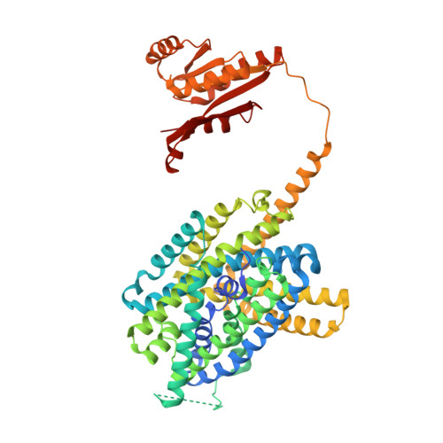
    Cyclic di-AMP is the only known essential second messenger in bacteria and archaea, regulating different proteins indispensable for numerous physiological processes. In particular, it controls various potassium and osmolyte transporters involved in osmoregulation. In Bacillus subtilis, the K + /H + symporter KimA of the KUP family is inactivated by c-di-AMP. KimA sustains survival at potassium limitation at low external pH by mediating potassium ion uptake. However, at elevated intracellular K + concentrations, further K + accumulation would be toxic. In this study, we reveal the molecular basis of how c-di-AMP binding inhibits KimA. We report cryo-EM structures of KimA with bound c-di-AMP in detergent solution and reconstituted in amphipols. By combining structural data with functional assays and molecular dynamics simulations we reveal how c-di-AMP modulates transport. We show that an intracellular loop in the transmembrane domain interacts with c-di-AMP bound to the adjacent cytosolic domain. This reduces the mobility of transmembrane helices at the cytosolic side of the K + binding site and therefore traps KimA in an inward-occluded conformation.


  • Organizational Affiliation

    Institute of Biochemistry, Goethe University Frankfurt, Frankfurt am Main, Germany.


Macromolecules
Find similar proteins by:  (by identity cutoff)  |  3D Structure
Entity ID: 1
MoleculeChains Sequence LengthOrganismDetailsImage
Potassium transporter KimA
A, B
607Bacillus subtilisMutation(s): 0 
Gene Names: kimAydaOBSU04320
Membrane Entity: Yes 
UniProt
Find proteins for P96589 (Bacillus subtilis (strain 168))
Explore P96589 
Go to UniProtKB:  P96589
Entity Groups  
Sequence Clusters30% Identity50% Identity70% Identity90% Identity95% Identity100% Identity
UniProt GroupP96589
Sequence Annotations
Expand
  • Reference Sequence
Small Molecules
Ligands 3 Unique
IDChains Name / Formula / InChI Key2D Diagram3D Interactions
2BA
Query on 2BA

Download Ideal Coordinates CCD File 
C [auth A],
K [auth B]
(2R,3R,3aS,5R,7aR,9R,10R,10aS,12R,14aR)-2,9-bis(6-amino-9H-purin-9-yl)octahydro-2H,7H-difuro[3,2-d:3',2'-j][1,3,7,9,2,8 ]tetraoxadiphosphacyclododecine-3,5,10,12-tetrol 5,12-dioxide
C20 H24 N10 O12 P2
PDXMFTWFFKBFIN-XPWFQUROSA-N
LMT
Query on LMT

Download Ideal Coordinates CCD File 
D [auth A]
E [auth A]
F [auth A]
G [auth A]
I [auth A]
D [auth A],
E [auth A],
F [auth A],
G [auth A],
I [auth A],
J [auth A],
L [auth B]
DODECYL-BETA-D-MALTOSIDE
C24 H46 O11
NLEBIOOXCVAHBD-QKMCSOCLSA-N
K
Query on K

Download Ideal Coordinates CCD File 
H [auth A],
M [auth B]
POTASSIUM ION
K
NPYPAHLBTDXSSS-UHFFFAOYSA-N
Experimental Data & Validation

Experimental Data

  • Method: ELECTRON MICROSCOPY
  • Resolution: 3.30 Å
  • Aggregation State: PARTICLE 
  • Reconstruction Method: SINGLE PARTICLE 
EM Software:
TaskSoftware PackageVersion
MODEL REFINEMENTPHENIX
RECONSTRUCTIONRELION3.1

Structure Validation

View Full Validation Report


Entry History & Funding Information

Deposition Data


Funding OrganizationLocationGrant Number
German Research Foundation (DFG)GermanyHA 6322/4-1
German Research Foundation (DFG)GermanyVO 1449/1-1

Revision History  (Full details and data files)

  • Version 1.0: 2023-01-18
    Type: Initial release
  • Version 2.0: 2023-05-17
    Type: Coordinate replacement
    Reason: Atoms with unrealistic or zero occupancies
    Changes: Advisory, Atomic model, Author supporting evidence, Data collection, Database references, Derived calculations, Polymer sequence, Refinement description, Source and taxonomy, Structure summary
  • Version 2.1: 2023-07-05
    Changes: Database references