8B43 | pdb_00008b43

Crystal structure of ferrioxamine transporter


Experimental Data Snapshot

  • Method: X-RAY DIFFRACTION
  • Resolution: 2.49 Å
  • R-Value Free: 
    0.259 (Depositor), 0.255 (DCC) 
  • R-Value Work: 
    0.217 (Depositor), 0.217 (DCC) 
  • R-Value Observed: 
    0.219 (Depositor) 

Starting Model: experimental
View more details

wwPDB Validation 3D Report Full Report

Validation slider image for 8B43

Ligand Structure Quality Assessment 


This is version 1.3 of the entry. See complete history

Literature

Interactions of TonB-dependent transporter FoxA with siderophores and antibiotics that affect binding, uptake, and signal transduction.

Chan, D.C.K.Josts, I.Koteva, K.Wright, G.D.Tidow, H.Burrows, L.L.

(2023) Proc Natl Acad Sci U S A 120: e2221253120-e2221253120

  • DOI: https://doi.org/10.1073/pnas.2221253120
  • Primary Citation Related Structures: 
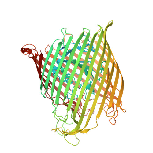
    8B43

  • PubMed Abstract: 

    The outer membrane of gram-negative bacteria prevents many antibiotics from reaching intracellular targets. However, some antimicrobials can take advantage of iron import transporters to cross this barrier. We showed previously that the thiopeptide antibiotic thiocillin exploits the nocardamine xenosiderophore transporter, FoxA, of the opportunistic pathogen Pseudomonas aeruginosa for uptake. Here, we show that FoxA also transports the xenosiderophore bisucaberin and describe at 2.5 Å resolution the crystal structure of bisucaberin bound to FoxA. Bisucaberin is distinct from other siderophores because it forms a 3:2 rather than 1:1 siderophore-iron complex. Mutations in a single extracellular loop of FoxA differentially affected nocardamine, thiocillin, and bisucaberin binding, uptake, and signal transduction. These results show that in addition to modulating ligand binding, the extracellular loops of siderophore transporters are of fundamental importance for controlling ligand uptake and its regulatory consequences, which have implications for the development of siderophore-antibiotic conjugates to treat difficult infections.


  • Organizational Affiliation
    • David Braley Center for Antibiotic Discovery, McMaster University, Hamilton, ON L8S 4K1, Canada.

Macromolecule Content 

  • Total Structure Weight: 92.63 kDa 
  • Atom Count: 5,457 
  • Modeled Residue Count: 677 
  • Deposited Residue Count: 820 
  • Unique protein chains: 1

Macromolecules

Find similar proteins by:|  3D Structure
Entity ID: 1
MoleculeChains  Sequence LengthOrganismDetailsImage
Metal-pseudopaline receptor CntO820Pseudomonas aeruginosaMutation(s): 0 
Gene Names: foxAPA2466
Membrane Entity: Yes 
UniProt
Find proteins for A0A0C7CQY7 (Pseudomonas aeruginosa)
Explore A0A0C7CQY7 
Go to UniProtKB:  A0A0C7CQY7
Entity Groups
Sequence Clusters30% Identity50% Identity70% Identity90% Identity95% Identity100% Identity
UniProt GroupA0A0C7CQY7
Sequence Annotations
Expand
Reference Sequence

Small Molecules

Ligands 4 Unique
IDChains Name / Formula / InChI Key2D Diagram3D Interactions
OX8
(Subject of Investigation/LOI)

Query on OX8


Download:Ideal Coordinates CCD File
J [auth A],
K [auth A],
L [auth A]
1,12-bis(oxidanyl)-1,6,12,17-tetrazacyclodocosane-2,5,13,16-tetrone
C18 H32 N4 O6
GTADQMQBQBOJIO-UHFFFAOYSA-N
BOG

Query on BOG


Download:Ideal Coordinates CCD File
E [auth A],
F [auth A],
G [auth A]
octyl beta-D-glucopyranoside
C14 H28 O6
HEGSGKPQLMEBJL-RKQHYHRCSA-N
SO4

Query on SO4


Download:Ideal Coordinates CCD File
B [auth A],
C [auth A],
D [auth A],
M [auth A]
SULFATE ION
O4 S
QAOWNCQODCNURD-UHFFFAOYSA-L
FE
(Subject of Investigation/LOI)

Query on FE


Download:Ideal Coordinates CCD File
H [auth A],
I [auth A]
FE (III) ION
Fe
VTLYFUHAOXGGBS-UHFFFAOYSA-N

Experimental Data & Validation

Experimental Data

  • Method: X-RAY DIFFRACTION
  • Resolution: 2.49 Å
  • R-Value Free:  0.259 (Depositor), 0.255 (DCC) 
  • R-Value Work:  0.217 (Depositor), 0.217 (DCC) 
  • R-Value Observed: 0.219 (Depositor) 
Space Group: P 32 2 1
Unit Cell:
Length ( Å )Angle ( ˚ )
a = 95.411α = 90
b = 95.411β = 90
c = 177.984γ = 120
Software Package:
Software NamePurpose
PHENIXrefinement
PDB_EXTRACTdata extraction
XDSdata reduction
Aimlessdata scaling
PHENIXphasing

Structure Validation

View Full Validation Report


Ligand Structure Quality Assessment 


Entry History 

& Funding Information

Deposition Data


Funding OrganizationLocationGrant Number
German Research Foundation (DFG)Germany--

Revision History  (Full details and data files)

  • Version 1.0: 2023-04-26
    Type: Initial release
  • Version 1.1: 2024-02-07
    Changes: Data collection, Refinement description
  • Version 1.2: 2024-11-13
    Changes: Structure summary
  • Version 1.3: 2026-01-21
    Changes: Database references, Source and taxonomy, Structure summary