8AVW

Cryo-EM structure of DrBphP in Pr state


Experimental Data Snapshot

  • Method: ELECTRON MICROSCOPY
  • Resolution: 3.62 Å
  • Aggregation State: PARTICLE 
  • Reconstruction Method: SINGLE PARTICLE 

wwPDB Validation   3D Report Full Report


This is version 1.1 of the entry. See complete history


Literature

Structural mechanism of signal transduction in a phytochrome histidine kinase.

Wahlgren, W.Y.Claesson, E.Tuure, I.Trillo-Muyo, S.Bodizs, S.Ihalainen, J.A.Takala, H.Westenhoff, S.

(2022) Nat Commun 13: 7673-7673

  • DOI: https://doi.org/10.1038/s41467-022-34893-3
  • Primary Citation of Related Structures:  
    8AVV, 8AVW, 8AVX

  • PubMed Abstract: 

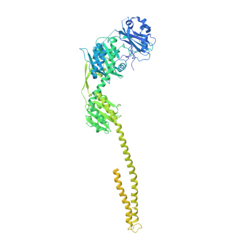
    Phytochrome proteins detect red/far-red light to guide the growth, motion, development and reproduction in plants, fungi, and bacteria. Bacterial phytochromes commonly function as an entrance signal in two-component sensory systems. Despite the availability of three-dimensional structures of phytochromes and other two-component proteins, the conformational changes, which lead to activation of the protein, are not understood. We reveal cryo electron microscopy structures of the complete phytochrome from Deinoccocus radiodurans in its resting and photoactivated states at 3.6 Å and 3.5 Å resolution, respectively. Upon photoactivation, the photosensory core module hardly changes its tertiary domain arrangement, but the connector helices between the photosensory and the histidine kinase modules open up like a zipper, causing asymmetry and disorder in the effector domains. The structures provide a framework for atom-scale understanding of signaling in phytochromes, visualize allosteric communication over several nanometers, and suggest that disorder in the dimeric arrangement of the effector domains is important for phosphatase activity in a two-component system. The results have implications for the development of optogenetic applications.


  • Organizational Affiliation

    Department of Chemistry and Molecular Biology, University of Gothenburg, Gothenburg, Sweden.


Macromolecules
Find similar proteins by:  (by identity cutoff)  |  3D Structure
Entity ID: 1
MoleculeChains Sequence LengthOrganismDetailsImage
Bacteriophytochrome,Response regulator
A, B
938Deinococcus radiodurans R1 = ATCC 13939 = DSM 20539Mutation(s): 0 
Gene Names: bphPDR_A0050DR_A0049
EC: 2.7.13.3
UniProt
Find proteins for Q9RZA4 (Deinococcus radiodurans (strain ATCC 13939 / DSM 20539 / JCM 16871 / CCUG 27074 / LMG 4051 / NBRC 15346 / NCIMB 9279 / VKM B-1422 / R1))
Explore Q9RZA4 
Go to UniProtKB:  Q9RZA4
Find proteins for Q9RZA5 (Deinococcus radiodurans (strain ATCC 13939 / DSM 20539 / JCM 16871 / CCUG 27074 / LMG 4051 / NBRC 15346 / NCIMB 9279 / VKM B-1422 / R1))
Explore Q9RZA5 
Go to UniProtKB:  Q9RZA5
Entity Groups  
Sequence Clusters30% Identity50% Identity70% Identity90% Identity95% Identity100% Identity
UniProt GroupsQ9RZA4Q9RZA5
Sequence Annotations
Expand
  • Reference Sequence
Small Molecules
Ligands 1 Unique
IDChains Name / Formula / InChI Key2D Diagram3D Interactions
LBV (Subject of Investigation/LOI)
Query on LBV

Download Ideal Coordinates CCD File 
C [auth A],
D [auth B]
3-[2-[(Z)-[3-(2-carboxyethyl)-5-[(Z)-(4-ethenyl-3-methyl-5-oxidanylidene-pyrrol-2-ylidene)methyl]-4-methyl-pyrrol-1-ium -2-ylidene]methyl]-5-[(Z)-[(3E)-3-ethylidene-4-methyl-5-oxidanylidene-pyrrolidin-2-ylidene]methyl]-4-methyl-1H-pyrrol-3- yl]propanoic acid
C33 H37 N4 O6
DKMLMZVDTGOEGU-ISEYCTJISA-O
Experimental Data & Validation

Experimental Data

  • Method: ELECTRON MICROSCOPY
  • Resolution: 3.62 Å
  • Aggregation State: PARTICLE 
  • Reconstruction Method: SINGLE PARTICLE 
EM Software:
TaskSoftware PackageVersion
MODEL REFINEMENTREFMAC5.8.0267
RECONSTRUCTIONcryoSPARC

Structure Validation

View Full Validation Report


Entry History & Funding Information

Deposition Data


Funding OrganizationLocationGrant Number
European Research Council (ERC)European Union725642
Academy of FinlandFinland330678

Revision History  (Full details and data files)

  • Version 1.0: 2022-12-21
    Type: Initial release
  • Version 1.1: 2022-12-28
    Changes: Database references