FtsEX-independent control of RipA-mediated cell separation in Corynebacteriales.
Gaday, Q., Megrian, D., Carloni, G., Martinez, M., Sokolova, B., Ben Assaya, M., Legrand, P., Brule, S., Haouz, A., Wehenkel, A.M., Alzari, P.M.(2022) Proc Natl Acad Sci U S A 119: e2214599119-e2214599119
- PubMed: 36469781 Search on PubMedSearch on PubMed Central
- DOI: https://doi.org/10.1073/pnas.2214599119
- Primary Citation Related Structures:
8AU6, 8AUC, 8AUD - PubMed Abstract:
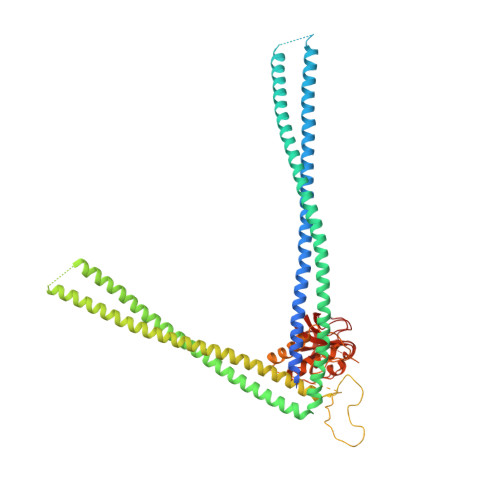
The bacterial cell wall is a multi-layered mesh, whose major component is peptidoglycan (PG), a sugar polymer cross-linked by short peptide stems. During cell division, a careful balance of PG synthesis and degradation, precisely coordinated both in time and space, is necessary to prevent uncontrolled destruction of the cell wall. In Corynebacteriales, the D,L endopeptidase RipA has emerged as a major PG hydrolase for cell separation, and RipA defaults have major implications for virulence of the human pathogens Mycobacterium tuberculosis and Corynebacterium diphtheriae . However, the precise mechanisms by which RipA mediates cell separation remain elusive. Here we report phylogenetic, biochemical, and structural analysis of the Corynebacterium glutamicum homologue of RipA, Cg1735. The crystal structures of full-length Cg1735 in two different crystal forms revealed the C-terminal NlpC/P60 catalytic domain obtruded by its N-terminal conserved coiled-coil domain, which locks the enzyme in an autoinhibited state. We show that this autoinhibition is relieved by the extracellular core domain of the transmembrane septal protein Cg1604. The crystal structure of Cg1604 revealed a (β/α) protein with an overall topology similar to that of receiver domains from response regulator proteins. The atomic model of the Cg1735-Cg1604 complex, based on bioinformatical and mutational analysis, indicates that a conserved, distal-membrane helical insertion in Cg1604 is responsible for Cg1735 activation. The reported data provide important insights into how intracellular cell division signal(s), yet to be identified, control PG hydrolysis during RipA-mediated cell separation in Corynebacteriales .
- Unité de Microbiologie Structurale, Institut Pasteur, CNRS, Université Paris Cité, Paris 75015, France.
Organizational Affiliation: